6913 results
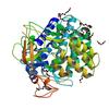
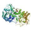
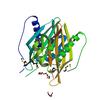
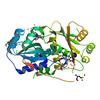
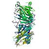
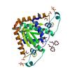
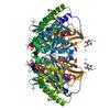
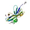
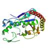
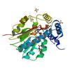
X-ray diffraction data for the Crystal structure of FMN-dependent nitroreductase BF3017 from Bacteroides fragilis NCTC 9343 (YP_212631.1) from Bacteroides fragilis NCTC 9343 at 1.55 A resolution
First author:
Joint Center for Structural Genomics (JCSG)
Resolution: 1.55 Å
R/Rfree: 0.16/0.19
Resolution: 1.55 Å
R/Rfree: 0.16/0.19
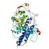
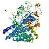
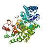
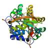
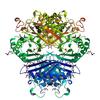
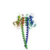
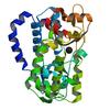
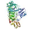
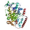
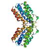
X-ray diffraction data for the Crystal structure of Putative antibiotic biosynthesis monooxygenase (NP_810307.1) from Bacteriodes thetaiotaomicron VPI-5482 at 1.30 A resolution
First author:
Joint Center for Structural Genomics (JCSG)
Resolution: 1.30 Å
R/Rfree: 0.14/0.17
Resolution: 1.30 Å
R/Rfree: 0.14/0.17
X-ray diffraction data for the Crystal structure of a glycerophosphodiester phosphodiesterase (BDI_0402) from Parabacteroides distasonis ATCC 8503 at 1.89 A resolution
First author:
Joint Center for Structural Genomics (JCSG)
Resolution: 1.89 Å
R/Rfree: 0.15/0.18
Resolution: 1.89 Å
R/Rfree: 0.15/0.18
X-ray diffraction data for the Crystal structure of Putative phosphatase (DUF442) (YP_001181608.1) from SHEWANELLA PUTREFACIENS CN-32 at 1.40 A resolution


First author:
Joint Center for Structural Genomics (JCSG)
Resolution: 1.40 Å
R/Rfree: 0.16/0.18
Resolution: 1.40 Å
R/Rfree: 0.16/0.18
X-ray diffraction data for the Crystal structure of Putative phosphatase (DUF442) (YP_001181608.1) from SHEWANELLA PUTREFACIENS CN-32 at 1.60 A resolution
First author:
Joint Center for Structural Genomics (JCSG)
Resolution: 1.60 Å
R/Rfree: 0.15/0.18
Resolution: 1.60 Å
R/Rfree: 0.15/0.18
X-ray diffraction data for the Crystal structure of a putative glycosylhydrolase (BT_3782) from Bacteroides thetaiotaomicron VPI-5482 at 1.89 A resolution
First author:
JOINT CENTER FOR STRUCTURAL GENOMICS (JCSG)
Resolution: 1.89 Å
R/Rfree: 0.15/0.19
Resolution: 1.89 Å
R/Rfree: 0.15/0.19
X-ray diffraction data for the Crystal structure of Putative glycosyl hydrolase (NP_813087.1) from BACTEROIDES THETAIOTAOMICRON VPI-5482 at 1.80 A resolution
First author:
Joint Center for Structural Genomics (JCSG)
Resolution: 1.80 Å
R/Rfree: 0.15/0.17
Resolution: 1.80 Å
R/Rfree: 0.15/0.17
X-ray diffraction data for the Crystal structure of Putative SnoaL-like polyketide cyclase (YP_509242.1) from Jannaschia Sp. CCS1 at 1.40 A resolution
First author:
Joint Center for Structural Genomics (JCSG)
Resolution: 1.40 Å
R/Rfree: 0.18/0.20
Resolution: 1.40 Å
R/Rfree: 0.18/0.20
X-ray diffraction data for the Crystal structure of Putative polyketide cyclase (NP_977253.1) from BACILLUS CEREUS ATCC 10987 at 1.91 A resolution
First author:
Joint Center for Structural Genomics (JCSG)
Resolution: 1.91 Å
R/Rfree: 0.17/0.22
Resolution: 1.91 Å
R/Rfree: 0.17/0.22
X-ray diffraction data for the CRYSTAL STRUCTURE OF A NLPC/P60 FAMILY PROTEIN (BCE_2878) FROM BACILLUS CEREUS ATCC 10987 AT 1.79 A RESOLUTION
First author:
Q. Xu
Resolution: 1.79 Å
R/Rfree: 0.16/0.20
Resolution: 1.79 Å
R/Rfree: 0.16/0.20
X-ray diffraction data for the Crystal structure of a Glycerophosphoryl diester phosphodiesterase (BDI_3922) from Parabacteroides distasonis ATCC 8503 at 1.55 A resolution
First author:
Joint Center for Structural Genomics (JCSG)
Resolution: 1.55 Å
R/Rfree: 0.17/0.20
Resolution: 1.55 Å
R/Rfree: 0.17/0.20
X-ray diffraction data for the Crystal structure of Putative glycerophosphoryl diester phosphodiesterase (NP_812074.1) from BACTEROIDES THETAIOTAOMICRON VPI-5482 at 1.35 A resolution
First author:
Joint Center for Structural Genomics (JCSG)
Resolution: 1.35 Å
R/Rfree: 0.15/0.17
Resolution: 1.35 Å
R/Rfree: 0.15/0.17
X-ray diffraction data for the Crystal structure of a branched chain amino acid ABC transporter periplasmic ligand-binding protein (Bxe_C0949) from BURKHOLDERIA XENOVORANS LB400 at 1.88 A resolution
First author:
Joint Center for Structural Genomics (JCSG)
Resolution: 1.88 Å
R/Rfree: 0.19/0.23
Resolution: 1.88 Å
R/Rfree: 0.19/0.23
X-ray diffraction data for the CRYSTAL STRUCTURE OF A PERIPLASMIC BINDING PROTEIN (BMA2936) FROM BURKHOLDERIA MALLEI AT 1.80 A RESOLUTION
First author:
Joint Center for Structural Genomics (JCSG)
Resolution: 1.80 Å
R/Rfree: 0.14/0.17
Resolution: 1.80 Å
R/Rfree: 0.14/0.17
X-ray diffraction data for the Crystal structure of Saro_0823 (YP_496102.1) a protein of unknown function from NOVOSPHINGOBIUM AROMATICIVORANS DSM 12444 at 1.22 A resolution
First author:
Joint Center for Structural Genomics (JCSG)
Resolution: 1.22 Å
R/Rfree: 0.12/0.15
Resolution: 1.22 Å
R/Rfree: 0.12/0.15
X-ray diffraction data for the CRYSTAL STRUCTURE OF AN UNCHARACTERIZED PROTEIN WITH A CYSTATIN-LIKE FOLD (CC_2572) FROM CAULOBACTER VIBRIOIDES AT 1.40 A RESOLUTION
First author:
Joint Center for Structural Genomics (JCSG)
Resolution: 1.40 Å
R/Rfree: 0.14/0.15
Resolution: 1.40 Å
R/Rfree: 0.14/0.15
X-ray diffraction data for the CRYSTAL STRUCTURE OF A SUSD SUPERFAMILY PROTEIN (BF_0597) FROM BACTEROIDES FRAGILIS AT 2.05 A RESOLUTION
First author:
Joint Center for Structural Genomics (JCSG)
Resolution: 2.05 Å
R/Rfree: 0.15/0.17
Resolution: 2.05 Å
R/Rfree: 0.15/0.17
X-ray diffraction data for the Crystal structure of SusD homolog (NP_809186.1) from BACTEROIDES THETAIOTAOMICRON VPI-5482 at 1.35 A resolution
First author:
Joint Center for Structural Genomics (JCSG)
Resolution: 1.35 Å
R/Rfree: 0.15/0.17
Resolution: 1.35 Å
R/Rfree: 0.15/0.17
X-ray diffraction data for the Crystal structure of Putative DNA-binding protein (YP_299413.1) from Ralstonia eutrophA JMP134 at 1.30 A resolution
First author:
Joint Center for Structural Genomics (JCSG)
Resolution: 1.30 Å
R/Rfree: 0.12/0.14
Resolution: 1.30 Å
R/Rfree: 0.12/0.14
X-ray diffraction data for the Crystal structure of putative ketosteroid isomerase (NP_103587.1) from Mesorhizobium loti at 1.45 A resolution
First author:
Joint Center for Structural Genomics (JCSG)
Resolution: 1.45 Å
R/Rfree: 0.15/0.19
Resolution: 1.45 Å
R/Rfree: 0.15/0.19
X-ray diffraction data for the Crystal structure of Endo-1,4-beta-xylanase (NP_811807.1) from BACTEROIDES THETAIOTAOMICRON VPI-5482 at 1.70 A resolution
First author:
Joint Center for Structural Genomics (JCSG)
Resolution: 1.70 Å
R/Rfree: 0.15/0.18
Resolution: 1.70 Å
R/Rfree: 0.15/0.18
X-ray diffraction data for the Crystal structure of Putative sugar isomerase. (YP_001305105.1) from Parabacteroides distasonis ATCC 8503 at 1.44 A resolution
First author:
Joint Center for Structural Genomics (JCSG)
Resolution: 1.44 Å
R/Rfree: 0.15/0.19
Resolution: 1.44 Å
R/Rfree: 0.15/0.19
X-ray diffraction data for the Crystal structure of a Sugar phosphate isomerase/epimerase (BDI_1903) from Parabacteroides distasonis ATCC 8503 at 1.85 A resolution
First author:
Joint Center for Structural Genomics (JCSG)
Resolution: 1.85 Å
R/Rfree: 0.19/0.24
Resolution: 1.85 Å
R/Rfree: 0.19/0.24
X-ray diffraction data for the Crystal structure of a Putative uncharacterized protein (BDI_1873) from Parabacteroides distasonis ATCC 8503 at 2.32 A resolution
First author:
Joint Center for Structural Genomics (JCSG)
Resolution: 2.32 Å
R/Rfree: 0.18/0.22
Resolution: 2.32 Å
R/Rfree: 0.18/0.22
X-ray diffraction data for the Crystal structure of Putative SnoaL-like polyketide cyclase (YP_563807.1) from SHEWANELLA DENITRIFICANS OS-217 at 1.70 A resolution
First author:
Joint Center for Structural Genomics (JCSG)
Resolution: 1.70 Å
R/Rfree: 0.19/0.24
Resolution: 1.70 Å
R/Rfree: 0.19/0.24
X-ray diffraction data for the Crystal structure of Putative histidinol-phosphate aminotransferase (YP_050345.1) from Erwinia carotovora atroseptica SCRI1043 at 1.80 A resolution
First author:
Joint Center for Structural Genomics (JCSG)
Resolution: 1.80 Å
R/Rfree: 0.18/0.21
Resolution: 1.80 Å
R/Rfree: 0.18/0.21
X-ray diffraction data for the Crystal structure of SusD superfamily protein (YP_001297730.1) from Bacteroides vulgatus ATCC 8482 at 1.55 A resolution
First author:
Joint Center for Structural Genomics (JCSG)
Resolution: 1.55 Å
R/Rfree: 0.17/0.21
Resolution: 1.55 Å
R/Rfree: 0.17/0.21
X-ray diffraction data for the Crystal structure of SusD-like carbohydrate binding protein (YP_001298396.1) from Bacteroides vulgatus ATCC 8482 at 1.70 A resolution
First author:
Joint Center for Structural Genomics (JCSG)
Resolution: 1.70 Å
R/Rfree: 0.15/0.17
Resolution: 1.70 Å
R/Rfree: 0.15/0.17
X-ray diffraction data for the Crystal structure of a PUTATIVE SUSD-LIKE CARBOHYDRATE BINDING PROTEIN (BT_1439) from BACTEROIDES THETAIOTAOMICRON VPI-5482 at 1.88 A resolution
First author:
Joint center for structural genomics (jcsg)
Resolution: 1.88 Å
R/Rfree: 0.17/0.21
Resolution: 1.88 Å
R/Rfree: 0.17/0.21
X-ray diffraction data for the Crystal structure of SusD superfamily protein (NP_809182.1) from BACTEROIDES THETAIOTAOMICRON VPI-5482 at 1.50 A resolution
First author:
Joint Center for Structural Genomics (JCSG)
Resolution: 1.50 Å
R/Rfree: 0.13/0.16
Resolution: 1.50 Å
R/Rfree: 0.13/0.16
X-ray diffraction data for the Crystal structure of a SusD superfamily protein (BDI_3964) from Parabacteroides distasonis ATCC 8503 at 1.95 A resolution

First author:
Joint Center for Structural Genomics (JCSG)
Resolution: 1.95 Å
R/Rfree: 0.14/0.19
Resolution: 1.95 Å
R/Rfree: 0.14/0.19
X-ray diffraction data for the Crystal structure of a SusD-like protein (BF3747) from Bacteroides fragilis NCTC 9343 at 2.70 A resolution
First author:
Joint Center for Structural Genomics (JCSG)
Resolution: 2.70 Å
R/Rfree: 0.18/0.21
Resolution: 2.70 Å
R/Rfree: 0.18/0.21
X-ray diffraction data for the CRYSTAL STRUCTURE OF A SUSD-LIKE CARBOHYDRATE BINDING PROTEIN (BF0978) FROM BACTEROIDES FRAGILIS NCTC 9343 AT 1.35 A RESOLUTION
First author:
Joint Center for Structural Genomics (JCSG)
Resolution: 1.35 Å
R/Rfree: 0.14/0.16
Resolution: 1.35 Å
R/Rfree: 0.14/0.16
X-ray diffraction data for the Crystal structure of Putative calcium/calmodulin dependent protein kinase II association domain (NP_636218.1) from XANTHOMONAS CAMPESTRIS at 1.70 A resolution
First author:
Joint Center for Structural Genomics (JCSG)
Resolution: 1.70 Å
R/Rfree: 0.18/0.22
Resolution: 1.70 Å
R/Rfree: 0.18/0.22
X-ray diffraction data for the Crystal structure of Putative sugar isomerase (YP_001305149.1) from Parabacteroides distasonis ATCC 8503 at 1.68 A resolution
First author:
Joint Center for Structural Genomics (JCSG)
Resolution: 1.68 Å
R/Rfree: 0.15/0.18
Resolution: 1.68 Å
R/Rfree: 0.15/0.18
X-ray diffraction data for the Crystal structure of Putative NADH dehydrogenase/NAD(P)H nitroreductase (BDI_1728) from Parabacteroides distasonis ATCC 8503 at 1.86 A resolution
First author:
Joint Center for Structural Genomics (JCSG)
Resolution: 1.86 Å
R/Rfree: 0.16/0.18
Resolution: 1.86 Å
R/Rfree: 0.16/0.18
X-ray diffraction data for the Crystal structure of Putative NADH dehydrogenase/NAD(P)H nitroreductase (NP_809094.1) from BACTEROIDES THETAIOTAOMICRON VPI-5482 at 1.54 A resolution
First author:
Joint Center for Structural Genomics (JCSG)
Resolution: 1.54 Å
R/Rfree: 0.15/0.18
Resolution: 1.54 Å
R/Rfree: 0.15/0.18
X-ray diffraction data for the Crystal Structure of Human Pseudouridylate Synthase 7
First author:
H. ZENG
Resolution: 2.26 Å
R/Rfree: 0.21/0.26
Resolution: 2.26 Å
R/Rfree: 0.21/0.26
X-ray diffraction data for the Crystal structure of a putative d-serine deaminase (bxe_a4060) from burkholderia xenovorans lb400 at 2.00 A resolution

First author:
Joint Center for Structural Genomics (JCSG)
Resolution: 2.00 Å
R/Rfree: 0.16/0.21
Resolution: 2.00 Å
R/Rfree: 0.16/0.21
X-ray diffraction data for the Crystal structure of a putative sulfurtransferase dsrE (Swol_2425) from Syntrophomonas wolfei str. Goettingen at 1.92 A resolution
First author:
Joint Center for Structural Genomics (JCSG)
Resolution: 1.92 Å
R/Rfree: 0.19/0.22
Resolution: 1.92 Å
R/Rfree: 0.19/0.22
X-ray diffraction data for the Crystal structure of putative hydrolase (YP_751971.1) from Shewanella frigidimarina NCIMB 400 at 1.80 A resolution
First author:
Joint Center for Structural Genomics (JCSG)
Resolution: 1.80 Å
R/Rfree: 0.17/0.21
Resolution: 1.80 Å
R/Rfree: 0.17/0.21
X-ray diffraction data for the Crystal structure of Putative glyoxalase superfamily protein (YP_299723.1) from RALSTONIA EUTROPHA JMP134 at 1.90 A resolution
First author:
Joint Center for Structural Genomics (JCSG)
Resolution: 1.90 Å
R/Rfree: 0.17/0.21
Resolution: 1.90 Å
R/Rfree: 0.17/0.21
X-ray diffraction data for the Crystal structure of a ftsz-like protein of unknown function (npun_r1471) from nostoc punctiforme pcc 73102 at 1.22 A resolution

First author:
Joint Center for Structural Genomics (JCSG)
Resolution: 1.22 Å
R/Rfree: 0.16/0.18
Resolution: 1.22 Å
R/Rfree: 0.16/0.18
X-ray diffraction data for the Crystal structure of a duf574 family protein (sav_2177) from streptomyces avermitilis ma-4680 at 1.80 A resolution
First author:
Joint Center for Structural Genomics (JCSG)
Resolution: 1.80 Å
R/Rfree: 0.20/0.25
Resolution: 1.80 Å
R/Rfree: 0.20/0.25
X-ray diffraction data for the CRYSTAL STRUCTURE OF a DUF574 family protein (SAV_2177) FROM STREPTOMYCES AVERMITILIS MA-4680 AT 1.45 A RESOLUTION


First author:
Joint Center for Structural Genomics (JCSG)
Resolution: 1.45 Å
R/Rfree: 0.16/0.18
Resolution: 1.45 Å
R/Rfree: 0.16/0.18
X-ray diffraction data for the Crystal structure of Protein with unknown function which belongs to Pfam DUF971 family (AFE_2189) from Acidithiobacillus ferrooxidans ATCC 23270 at 1.93 A resolution

First author:
Joint Center for Structural Genomics (JCSG)
Resolution: 1.93 Å
R/Rfree: 0.20/0.22
Resolution: 1.93 Å
R/Rfree: 0.20/0.22
X-ray diffraction data for the Crystal structure of a putative 3-keto-5-aminohexanoate cleavage enzyme (reut_c6226) from ralstonia eutropha jmp134 at 1.72 A resolution

First author:
Joint Center for Structural Genomics (JCSG)
Resolution: 1.72 Å
R/Rfree: 0.15/0.18
Resolution: 1.72 Å
R/Rfree: 0.15/0.18
X-ray diffraction data for the Crystal structure of a prokaryotic domain of unknown function (duf849) with a tim barrel fold (bxe_c0966) from burkholderia xenovorans lb400 at 1.75 A resolution
First author:
Joint Center for Structural Genomics (JCSG)
Resolution: 1.75 Å
R/Rfree: 0.18/0.22
Resolution: 1.75 Å
R/Rfree: 0.18/0.22
X-ray diffraction data for the Crystal structure of a duf849 family protein (bxe_c0271) from burkholderia xenovorans lb400 at 1.90 A resolution
First author:
Joint Center for Structural Genomics (JCSG)
Resolution: 1.90 Å
R/Rfree: 0.16/0.18
Resolution: 1.90 Å
R/Rfree: 0.16/0.18
X-ray diffraction data for the Crystal structure of PROTEIN OF UNKNOWN FUNCTION (NP_070038.1) from ARCHAEOGLOBUS FULGIDUS at 1.89 A resolution
First author:
Joint Center for Structural Genomics (JCSG)
Resolution: 1.89 Å
R/Rfree: 0.16/0.21
Resolution: 1.89 Å
R/Rfree: 0.16/0.21
X-ray diffraction data for the Crystal structure of pleckstrin homology domain (YP_926556.1) from SHEWANELLA AMAZONENSIS SB2B at 1.99 A resolution
First author:
Q. Xu
Resolution: 1.99 Å
R/Rfree: 0.19/0.23
Resolution: 1.99 Å
R/Rfree: 0.19/0.23
X-ray diffraction data for the Crystal structure of a duf1185 family protein (bb2672) from bordetella bronchiseptica rb50 at 1.70 A resolution
First author:
C. Bakolitsa
Resolution: 1.70 Å
R/Rfree: 0.14/0.17
Resolution: 1.70 Å
R/Rfree: 0.14/0.17
X-ray diffraction data for the Crystal structure of a nifx-associated protein of unknown function (afe_1514) from acidithiobacillus ferrooxidans atcc at 2.00 A resolution
First author:
Joint Center for Structural Genomics (JCSG)
Resolution: 2.00 Å
R/Rfree: 0.20/0.25
Resolution: 2.00 Å
R/Rfree: 0.20/0.25
X-ray diffraction data for the CRYSTAL STRUCTURE OF a GAF domain containing protein that belongs to Pfam DUF484 family (PA5279) FROM PSEUDOMONAS AERUGINOSA AT 2.43 A RESOLUTION
First author:
Joint Center for Structural Genomics (JCSG)
Resolution: 2.43 Å
R/Rfree: 0.21/0.27
Resolution: 2.43 Å
R/Rfree: 0.21/0.27
X-ray diffraction data for the Crystal structure of Putative SAM Dependent Methyltransferase in Complex with SAH (NP_744700.1) from PSEUDOMONAS PUTIDA KT2440 at 2.10 A resolution
First author:
Joint Center for Structural Genomics (JCSG)
Resolution: 2.10 Å
R/Rfree: 0.16/0.18
Resolution: 2.10 Å
R/Rfree: 0.16/0.18
X-ray diffraction data for the Crystal structure of DsrE/DsrF-like family protein (NP_342589.1) from SULFOLOBUS SOLFATARICUS at 1.49 A resolution
First author:
Joint Center for Structural Genomics (JCSG)
Resolution: 1.49 Å
R/Rfree: 0.12/0.14
Resolution: 1.49 Å
R/Rfree: 0.12/0.14
X-ray diffraction data for the Crystal structure of Putative gamma-glutamylcysteine synthetase (YP_546622.1) from METHYLOBACILLUS FLAGELLATUS KT at 1.90 A resolution
First author:
Joint Center for Structural Genomics (JCSG)
Resolution: 1.90 Å
R/Rfree: 0.15/0.16
Resolution: 1.90 Å
R/Rfree: 0.15/0.16
X-ray diffraction data for the Crystal structure of novel protein of unknown function (YP_111841.1) from BURKHOLDERIA PSEUDOMALLEI K96243 at 1.90 A resolution
First author:
Joint Center for Structural Genomics (JCSG)
Resolution: 1.90 Å
R/Rfree: 0.18/0.22
Resolution: 1.90 Å
R/Rfree: 0.18/0.22
X-ray diffraction data for the Crystal structure of Putative metallopeptidase (YP_676511.1) from MESORHIZOBIUM SP. BNC1 at 2.13 A resolution


First author:
Joint Center for Structural Genomics (JCSG)
Resolution: 2.13 Å
R/Rfree: 0.18/0.23
Resolution: 2.13 Å
R/Rfree: 0.18/0.23
X-ray diffraction data for the Crystal structure of acetoacetate decarboxylase (YP_001047042.1) from Methanoculleus marisnigri JR1 at 1.60 A resolution
First author:
Joint Center for Structural Genomics (JCSG)
Resolution: 1.60 Å
R/Rfree: 0.23/0.26
Resolution: 1.60 Å
R/Rfree: 0.23/0.26
X-ray diffraction data for the Crystal structure of Structural Genomics, unknown function (YP_766765.1) from Rhizobium leguminosarum BV. viciae 3841 at 1.40 A resolution
First author:
Joint Center for Structural Genomics (JCSG)
Resolution: 1.40 Å
R/Rfree: 0.18/0.19
Resolution: 1.40 Å
R/Rfree: 0.18/0.19
X-ray diffraction data for the CRYSTAL STRUCTURE OF a DUF849 family protein (PDEN_3495) FROM PARACOCCUS DENITRIFICANS PD1222 AT 1.90 A RESOLUTION
First author:
Joint Center for Structural Genomics (JCSG)
Resolution: 1.90 Å
R/Rfree: 0.21/0.25
Resolution: 1.90 Å
R/Rfree: 0.21/0.25
X-ray diffraction data for the Crystal structure of prophage tail protein gp18 (NP_465809.1) from Listeria monocytogenes EGD-e at 1.70 A resolution
First author:
Joint Center for Structural Genomics (JCSG)
Resolution: 1.70 Å
R/Rfree: 0.19/0.22
Resolution: 1.70 Å
R/Rfree: 0.19/0.22
X-ray diffraction data for the Crystal structure of acetoacetate decarboxylase (ADC) (YP_094708.1) from Legionella pneumophila subsp. pneumophila str. Philadelphia 1 at 1.60 A resolution
First author:
Joint Center for Structural Genomics (JCSG)
Resolution: 1.60 Å
R/Rfree: 0.19/0.23
Resolution: 1.60 Å
R/Rfree: 0.19/0.23
X-ray diffraction data for the Crystal structure of a predicted zincin-like metalloprotease (acel_2062) from acidothermus cellulolyticus 11b at 1.80 A resolution
First author:
Joint Center for Structural Genomics (JCSG)
Resolution: 1.80 Å
R/Rfree: 0.18/0.20
Resolution: 1.80 Å
R/Rfree: 0.18/0.20
X-ray diffraction data for the CRYSTAL STRUCTURE OF A PROTEIN OF UNKNOWN FUNCTION FROM DUF427 FAMILY (RSPH17029_0682) FROM RHODOBACTER SPHAEROIDES 2.4.1 AT 2.51 A RESOLUTION
First author:
Joint Center for Structural Genomics (JCSG)
Resolution: 2.51 Å
R/Rfree: 0.21/0.23
Resolution: 2.51 Å
R/Rfree: 0.21/0.23
X-ray diffraction data for the Crystal structure of a duf416 family protein (maqu_0942) from marinobacter aquaeolei vt8 at 2.00 A resolution


First author:
Joint Center for Structural Genomics (JCSG)
Resolution: 2.00 Å
R/Rfree: 0.17/0.20
Resolution: 2.00 Å
R/Rfree: 0.17/0.20
X-ray diffraction data for the Crystal Structure of Wild-Type Human Prolidase with GlyPro ligand
First author:
P. Wilk
Resolution: 1.55 Å
R/Rfree: 0.15/0.17
Resolution: 1.55 Å
R/Rfree: 0.15/0.17
X-ray diffraction data for the Crystal Structure of Wild-Type Human Prolidase with Mn ions and Pro ligand
First author:
P. Wilk
Resolution: 1.73 Å
R/Rfree: 0.15/0.18
Resolution: 1.73 Å
R/Rfree: 0.15/0.18
X-ray diffraction data for the Crystal Structure of Wild-Type Human Prolidase with Mn ions
First author:
P. Wilk
Resolution: 1.48 Å
R/Rfree: 0.15/0.17
Resolution: 1.48 Å
R/Rfree: 0.15/0.17
X-ray diffraction data for the Crystal Structure of Wild-Type Human Prolidase with Mg ions and LeuPro ligand
First author:
P. Wilk
Resolution: 1.49 Å
R/Rfree: 0.15/0.17
Resolution: 1.49 Å
R/Rfree: 0.15/0.17
X-ray diffraction data for the Crystal Structure of Ser202Phe mutant of Human Prolidase with Mn ions and GlyPro ligand
First author:
P. Wilk
Resolution: 1.50 Å
R/Rfree: 0.15/0.18
Resolution: 1.50 Å
R/Rfree: 0.15/0.18
X-ray diffraction data for the Crystal Structure of Arg184Gln mutant of Human Prolidase with Mn ions and GlyPro ligand
First author:
P. Wilk
Resolution: 1.55 Å
R/Rfree: 0.16/0.17
Resolution: 1.55 Å
R/Rfree: 0.16/0.17
X-ray diffraction data for the Crystal Structure of Arg184Gln mutant of Human Prolidase with Mn ions and LeuPro ligand

First author:
P. Wilk
Resolution: 1.78 Å
R/Rfree: 0.15/0.18
Resolution: 1.78 Å
R/Rfree: 0.15/0.18
X-ray diffraction data for the Crystal Structure of Arg184Gln mutant of Human Prolidase with Mn ions and Cacodylate ligand
First author:
P. Wilk
Resolution: 1.48 Å
R/Rfree: 0.15/0.17
Resolution: 1.48 Å
R/Rfree: 0.15/0.17
X-ray diffraction data for the Crystal Structure of Gly448Arg mutant of Human Prolidase with Mn ions and GlyPro ligand
First author:
P. Wilk
Resolution: 1.80 Å
R/Rfree: 0.16/0.19
Resolution: 1.80 Å
R/Rfree: 0.16/0.19
X-ray diffraction data for the Crystal Structure of Gly278Asp mutant of Human Prolidase with Mn ions and GlyPro ligand
First author:
P. Wilk
Resolution: 1.70 Å
R/Rfree: 0.14/0.17
Resolution: 1.70 Å
R/Rfree: 0.14/0.17
X-ray diffraction data for the Crystal Structure of delTyr231 mutant of Human Prolidase with Mn ions and GlyPro ligand
First author:
P. Wilk
Resolution: 1.56 Å
R/Rfree: 0.12/0.15
Resolution: 1.56 Å
R/Rfree: 0.12/0.15
X-ray diffraction data for the Crystal Structure of delGlu452 mutant of Human Prolidase with Mn ions and GlyPro ligand
First author:
P. Wilk
Resolution: 1.90 Å
R/Rfree: 0.15/0.18
Resolution: 1.90 Å
R/Rfree: 0.15/0.18
X-ray diffraction data for the Crystal Structure of Asp276Asn mutant of Human Prolidase with Mn ions and GlyPro ligand
First author:
P. Wilk
Resolution: 1.43 Å
R/Rfree: 0.13/0.16
Resolution: 1.43 Å
R/Rfree: 0.13/0.16
X-ray diffraction data for the Crystal structure of an ala racemase-like protein (il1761) from idiomarina loihiensis at 1.50 A resolution
First author:
Joint Center for Structural Genomics (JCSG)
Resolution: 1.50 Å
R/Rfree: 0.14/0.16
Resolution: 1.50 Å
R/Rfree: 0.14/0.16
X-ray diffraction data for the Crystal structure of a DinB-like protein (CPS_3021) from Colwellia psychrerythraea 34H at 2.20 A resolution
First author:
Joint Center for Structural Genomics (JCSG)
Resolution: 2.20 Å
R/Rfree: 0.18/0.21
Resolution: 2.20 Å
R/Rfree: 0.18/0.21
X-ray diffraction data for the Crystal structure of Putative hydrolase (YP_002548124.1) from Agrobacterium vitis S4 at 1.80 A resolution
First author:
Joint Center for Structural Genomics (JCSG)
Resolution: 1.80 Å
R/Rfree: 0.16/0.19
Resolution: 1.80 Å
R/Rfree: 0.16/0.19
X-ray diffraction data for the CRYSTAL STRUCTURE OF A PHENYLACETATE-COA OXYGENASE SUBUNIT PAAB (REUT_A2307) FROM RALSTONIA EUTROPHA JMP134 AT 2.65 A RESOLUTION
First author:
Joint Center for Structural Genomics (JCSG)
Resolution: 2.65 Å
R/Rfree: 0.19/0.23
Resolution: 2.65 Å
R/Rfree: 0.19/0.23
X-ray diffraction data for the Crystal structure of a Putative Metal-dependent Hydrolase (YP_001336084.1) from Klebsiella pneumoniae subsp. pneumoniae MGH 78578 at 1.95 A resolution
First author:
Q. Xu
Resolution: 1.95 Å
R/Rfree: 0.18/0.21
Resolution: 1.95 Å
R/Rfree: 0.18/0.21
X-ray diffraction data for the Crystal structure of a Putative Metal-dependent Hydrolase (YP_001336084.1) from Klebsiella pneumoniae subsp. pneumoniae MGH 78578 at 2.20 A resolution


First author:
Q. Xu
Resolution: 2.20 Å
R/Rfree: 0.17/0.20
Resolution: 2.20 Å
R/Rfree: 0.17/0.20
X-ray diffraction data for the Crystal structure of a dinb-like protein (yjoa, bsu12410) from bacillus subtilis at 2.30 A resolution
First author:
Joint Center for Structural Genomics (JCSG)
Resolution: 2.30 Å
R/Rfree: 0.22/0.27
Resolution: 2.30 Å
R/Rfree: 0.22/0.27
X-ray diffraction data for the Crystal structure of NtrC-like two-domain protein (RER070207001320) from Eubacterium rectale at 2.10 A resolution
First author:
Q. Xu
Resolution: 2.10 Å
R/Rfree: 0.19/0.22
Resolution: 2.10 Å
R/Rfree: 0.19/0.22
X-ray diffraction data for the Crystal structure of a multidomain protein with nucleic acid binding domains (sp_0946) from streptococcus pneumoniae tigr4 at 1.40 A resolution

First author:
Y. Matsumoto
Resolution: 1.40 Å
R/Rfree: 0.15/0.18
Resolution: 1.40 Å
R/Rfree: 0.15/0.18
X-ray diffraction data for the Crystal structure of a duf1470 family protein (jann_2411) from jannaschia sp. ccs1 at 1.45 A resolution
First author:
C. Bakolitsa
Resolution: 1.45 Å
R/Rfree: 0.14/0.16
Resolution: 1.45 Å
R/Rfree: 0.14/0.16
X-ray diffraction data for the CRYSTAL STRUCTURE OF a DUF1285 family protein (SPO_0140) FROM SILICIBACTER POMEROYI DSS-3 AT 2.50 A RESOLUTION
First author:
G.W. Han
Resolution: 2.50 Å
R/Rfree: 0.22/0.26
Resolution: 2.50 Å
R/Rfree: 0.22/0.26
X-ray diffraction data for the Crystal structure of a duf1185 family protein (spo0826) from silicibacter pomeroyi dss-3 at 2.10 A resolution
First author:
C. Bakolitsa
Resolution: 2.10 Å
R/Rfree: 0.20/0.26
Resolution: 2.10 Å
R/Rfree: 0.20/0.26
X-ray diffraction data for the CRYSTAL STRUCTURE OF a prokaryotic domain of unknown function (DUF849) member (SPOA0042) FROM SILICIBACTER POMEROYI DSS-3 AT 1.45 A RESOLUTION
First author:
Joint Center for Structural Genomics (JCSG)
Resolution: 1.45 Å
R/Rfree: 0.13/0.16
Resolution: 1.45 Å
R/Rfree: 0.13/0.16
X-ray diffraction data for the Crystal structure of a dinb-like protein (bce_4655) from bacillus cereus atcc 10987 at 2.01 A resolution


First author:
Joint Center for Structural Genomics (JCSG)
Resolution: 2.01 Å
R/Rfree: 0.23/0.27
Resolution: 2.01 Å
R/Rfree: 0.23/0.27
X-ray diffraction data for the Crystal structure of a putative metal-dependent hydrolase (yiza, bsu10800) from bacillus subtilis at 1.90 A resolution
First author:
Joint Center for Structural Genomics (JCSG)
Resolution: 1.90 Å
R/Rfree: 0.19/0.23
Resolution: 1.90 Å
R/Rfree: 0.19/0.23
X-ray diffraction data for the Crystal structure of a duf692 family protein (hs_1138) from haemophilus somnus 129pt at 2.20 A resolution


First author:
Joint Center for Structural Genomics (JCSG)
Resolution: 2.20 Å
R/Rfree: 0.20/0.26
Resolution: 2.20 Å
R/Rfree: 0.20/0.26
X-ray diffraction data for the Crystal structure of N-formylglutamate amidohydrolase (YP_297560.1) from Ralstonia eutropha JMP134 at 2.00 A resolution
First author:
Joint Center for Structural Genomics (JCSG)
Resolution: 2.00 Å
R/Rfree: 0.20/0.25
Resolution: 2.00 Å
R/Rfree: 0.20/0.25
X-ray diffraction data for the Crystal structure of tryptophan halogenase (YP_750003.1) from Shewanella frigidimarina NCIMB 400 at 1.50 A resolution
First author:
Joint Center for Structural Genomics (JCSG)
Resolution: 1.50 Å
R/Rfree: 0.15/0.18
Resolution: 1.50 Å
R/Rfree: 0.15/0.18
X-ray diffraction data for the CRYSTAL STRUCTURE OF A PUTATIVE METHYLTRANSFERASE (TFU_2867) FROM THERMOBIFIDA FUSCA YX AT 1.95 A RESOLUTION
First author:
Joint Center for Structural Genomics (JCSG)
Resolution: 1.95 Å
R/Rfree: 0.17/0.21
Resolution: 1.95 Å
R/Rfree: 0.17/0.21
X-ray diffraction data for the CRYSTAL STRUCTURE OF a DUF416 family protein (SBAL_3149) FROM SHEWANELLA BALTICA OS155 AT 1.91 A RESOLUTION
First author:
Joint Center for Structural Genomics (JCSG)
Resolution: 1.91 Å
R/Rfree: 0.17/0.22
Resolution: 1.91 Å
R/Rfree: 0.17/0.22