6913 results
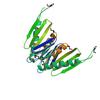
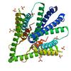
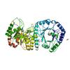
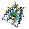
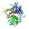
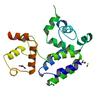
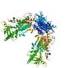
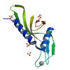
X-ray diffraction data for the Crystal structure of a Putative glutathionylspermidine synthase (Mfla_0391) from METHYLOBACILLUS FLAGELLATUS KT at 2.35 A resolution
First author:
Joint Center for Structural Genomics (JCSG)
Resolution: 2.35 Å
R/Rfree: 0.18/0.20
Resolution: 2.35 Å
R/Rfree: 0.18/0.20
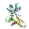
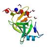
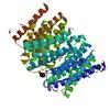
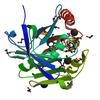
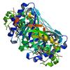
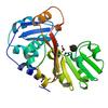
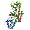
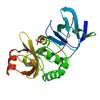
X-ray diffraction data for the Crystal structure of predicted acetamidase/formamidase (YP_546212.1) from Methylobacillus flagellatus KT at 1.58 A resolution
First author:
Joint Center for Structural Genomics (JCSG)
Resolution: 1.58 Å
R/Rfree: 0.14/0.18
Resolution: 1.58 Å
R/Rfree: 0.14/0.18
X-ray diffraction data for the CRYSTAL STRUCTURE OF A FIC FAMILY PROTEIN (SO_4266) FROM SHEWANELLA ONEIDENSIS AT 1.6 A RESOLUTION
First author:
D. Das
Resolution: 1.60 Å
R/Rfree: 0.17/0.20
Resolution: 1.60 Å
R/Rfree: 0.17/0.20
X-ray diffraction data for the CRYSTAL STRUCTURE OF A PUTATIVE PREPHENATE DEHYDROGENASE (CGL0226) FROM CORYNEBACTERIUM GLUTAMICUM ATCC 13032 AT 2.60 A RESOLUTION
First author:
Joint Center for Structural Genomics (JCSG)
Resolution: 2.60 Å
R/Rfree: 0.21/0.25
Resolution: 2.60 Å
R/Rfree: 0.21/0.25
X-ray diffraction data for the CRYSTAL STRUCTURE OF A PHAGE-RELATED PROTEIN (BB3626) FROM BORDETELLA BRONCHISEPTICA RB50 AT 2.05 A RESOLUTION
First author:
Joint Center for Structural Genomics (JCSG)
Resolution: 2.05 Å
R/Rfree: 0.19/0.23
Resolution: 2.05 Å
R/Rfree: 0.19/0.23
X-ray diffraction data for the Crystal structure of a putative dna binding protein (ape_0880a) from aeropyrum pernix k1 at 2.21 A resolution
First author:
Joint Center for Structural Genomics (JCSG)
Resolution: 2.21 Å
R/Rfree: 0.20/0.26
Resolution: 2.21 Å
R/Rfree: 0.20/0.26
X-ray diffraction data for the CRYSTAL STRUCTURE OF AN UNCHARACTERIZED PROTEIN FROM DUF3069 FAMILY (SAMA_2622) FROM SHEWANELLA AMAZONENSIS SB2B AT 1.95 A RESOLUTION
First author:
Joint Center for Structural Genomics (JCSG)
Resolution: 1.95 Å
R/Rfree: 0.21/0.25
Resolution: 1.95 Å
R/Rfree: 0.21/0.25
X-ray diffraction data for the Crystal structure of a duf2853 member protein (jann_4075) from jannaschia sp. ccs1 at 1.500 A resolution
First author:
Joint Center for Structural Genomics (JCSG)
Resolution: 1.50 Å
R/Rfree: 0.18/0.20
Resolution: 1.50 Å
R/Rfree: 0.18/0.20
X-ray diffraction data for the Crystal structure of uncharacterized protein (YP_563039.1) from Shewanella denitrificans OS217 at 1.80 A resolution
First author:
Joint Center for Structural Genomics (JCSG)
Resolution: 1.80 Å
R/Rfree: 0.17/0.18
Resolution: 1.80 Å
R/Rfree: 0.17/0.18
X-ray diffraction data for the CRYSTAL STRUCTURE OF a putative oxidoreductase of the DsrE/DsrF-like family (SSO1126) FROM SULFOLOBUS SOLFATARICUS P2 AT 2.09 A RESOLUTION
First author:
Joint Center for Structural Genomics (JCSG)
Resolution: 2.09 Å
R/Rfree: 0.18/0.20
Resolution: 2.09 Å
R/Rfree: 0.18/0.20
X-ray diffraction data for the CRYSTAL STRUCTURE OF A PUTATIVE HYDROLASE OF THE ALPHA/BETA SUPERFAMILY (XCC1541) FROM XANTHOMONAS CAMPESTRIS PV. CAMPESTRIS AT 1.35 A RESOLUTION
First author:
Joint Center for Structural Genomics (JCSG)
Resolution: 1.35 Å
R/Rfree: 0.18/0.22
Resolution: 1.35 Å
R/Rfree: 0.18/0.22
X-ray diffraction data for the Crystal structure of S-adenosylmethionine-dependent methyltransferase (NP_349143.1) from Clostridium acetobutylicum at 2.00 A resolution
First author:
Joint Center for Structural Genomics (JCSG)
Resolution: 2.00 Å
R/Rfree: 0.17/0.20
Resolution: 2.00 Å
R/Rfree: 0.17/0.20
X-ray diffraction data for the CRYSTAL STRUCTURE OF A PUTATIVE LIPID BINDING PROTEIN (GSU0061) FROM GEOBACTER SULFURREDUCENS PCA AT 1.80 A RESOLUTION
First author:
Joint Center for Structural Genomics (JCSG)
Resolution: 1.80 Å
R/Rfree: 0.19/0.22
Resolution: 1.80 Å
R/Rfree: 0.19/0.22
X-ray diffraction data for the Crystal structure of a methyltransferase (NP_951602.1) from Geobacter sulfurreducens at 1.90 A resolution

First author:
Joint Center for Structural Genomics (JCSG)
Resolution: 1.90 Å
R/Rfree: 0.20/0.23
Resolution: 1.90 Å
R/Rfree: 0.20/0.23
X-ray diffraction data for the CRYSTAL STRUCTURE OF A PROTEIN WITH UNKNOWN FUNCTION FROM DUF3224 FAMILY (SO_1590) FROM SHEWANELLA ONEIDENSIS MR-1 AT 1.84 A RESOLUTION
First author:
Joint Center for Structural Genomics (JCSG)
Resolution: 1.84 Å
R/Rfree: 0.18/0.21
Resolution: 1.84 Å
R/Rfree: 0.18/0.21
X-ray diffraction data for the Crystal structure of a protein with unknown function (YP_749275.1) from Shewanella Frigidimarina NCIMB 400 at 1.80 A resolution
First author:
A. Kumar
Resolution: 1.80 Å
R/Rfree: 0.18/0.23
Resolution: 1.80 Å
R/Rfree: 0.18/0.23
X-ray diffraction data for the CRYSTAL STRUCTURE OF AN UNCHARACTERIZED PROTEIN FROM DUF3478 FAMILY WITH A SPOIIAA-LIKE FOLD (SHEW_3102) FROM SHEWANELLA LOIHICA PV-4 AT 2.25 A RESOLUTION
First author:
A. Kumar
Resolution: 2.25 Å
R/Rfree: 0.18/0.24
Resolution: 2.25 Å
R/Rfree: 0.18/0.24
X-ray diffraction data for the Crystal structure of a cystatin-like fold protein (bxe_b1374) from burkholderia xenovorans lb400 at 2.00 A resolution

First author:
Joint Center for Structural Genomics (JCSG)
Resolution: 2.00 Å
R/Rfree: 0.19/0.24
Resolution: 2.00 Å
R/Rfree: 0.19/0.24
X-ray diffraction data for the CRYSTAL STRUCTURE OF A PROTEIN WITH UNKNOWN FUNCTION FROM DUF3225 FAMILY (ECA3500) FROM PECTOBACTERIUM ATROSEPTICUM SCRI1043 AT 2.32 A RESOLUTION
First author:
Joint Center for Structural Genomics (JCSG)
Resolution: 2.32 Å
R/Rfree: 0.19/0.24
Resolution: 2.32 Å
R/Rfree: 0.19/0.24
X-ray diffraction data for the Crystal structure of TehB-like SAM-dependent methyltransferase (NP_600671.1) from Corynebacterium glutamicum ATCC 13032 Kitasato at 2.00 A resolution
First author:
Joint Center for Structural Genomics (JCSG)
Resolution: 2.00 Å
R/Rfree: 0.17/0.21
Resolution: 2.00 Å
R/Rfree: 0.17/0.21
X-ray diffraction data for the CRYSTAL STRUCTURE OF A PUTATIVE ATPASE (CHU_0153) FROM CYTOPHAGA HUTCHINSONII ATCC 33406 AT 2.00 A RESOLUTION
First author:
Joint Center for Structural Genomics (JCSG)
Resolution: 2.00 Å
R/Rfree: 0.18/0.22
Resolution: 2.00 Å
R/Rfree: 0.18/0.22
X-ray diffraction data for the Crystal structure of a sec-c motif containing protein (psyc_2064) from psychrobacter arcticus at 1.75 A resolution
First author:
Joint Center for Structural Genomics (JCSG)
Resolution: 1.75 Å
R/Rfree: 0.19/0.24
Resolution: 1.75 Å
R/Rfree: 0.19/0.24
X-ray diffraction data for the CRYSTAL STRUCTURE OF A PUTATIVE PHOSPHONOPYRUVATE HYDROLASE (MLL9387) FROM MESORHIZOBIUM LOTI MAFF303099 AT 2.15 A RESOLUTION
First author:
Joint Center for Structural Genomics (JCSG)
Resolution: 2.15 Å
R/Rfree: 0.16/0.20
Resolution: 2.15 Å
R/Rfree: 0.16/0.20
X-ray diffraction data for the Crystal structure of a dinb family member protein (sden_0562) from shewanella denitrificans at 1.83 A resolution
First author:
Joint Center for Structural Genomics (JCSG)
Resolution: 1.83 Å
R/Rfree: 0.16/0.20
Resolution: 1.83 Å
R/Rfree: 0.16/0.20
X-ray diffraction data for the Crystal structure of putative tail lysozyme (NP_952040.1) from GEOBACTER SULFURREDUCENS at 1.44 A resolution
First author:
Joint Center for Structural Genomics (JCSG)
Resolution: 1.44 Å
R/Rfree: 0.17/0.20
Resolution: 1.44 Å
R/Rfree: 0.17/0.20
X-ray diffraction data for the Crystal structure of a putative dioxygenase (npun_f1925) from nostoc punctiforme pcc 73102 at 1.46 A resolution
First author:
Joint Center for Structural Genomics (JCSG)
Resolution: 1.46 Å
R/Rfree: 0.19/0.21
Resolution: 1.46 Å
R/Rfree: 0.19/0.21
X-ray diffraction data for the Crystal structure of putative dioxygenase (YP_555069.1) from Burkholderia xenovorans LB400 at 1.40 A resolution

First author:
Joint Center for Structural Genomics (JCSG)
Resolution: 1.40 Å
R/Rfree: 0.14/0.17
Resolution: 1.40 Å
R/Rfree: 0.14/0.17
X-ray diffraction data for the Crystal structure of putative dioxygenase (YP_555069.1) from Burkholderia Xenovorans LB400 at 1.70 A resolution
First author:
Joint Center for Structural Genomics (JCSG)
Resolution: 1.70 Å
R/Rfree: 0.18/0.22
Resolution: 1.70 Å
R/Rfree: 0.18/0.22
X-ray diffraction data for the Crystal structure of XisI protein-like (YP_323822.1) from Anabaena Variabilis ATCC 29413 at 1.85 A resolution
First author:
W.C. Hwang
Resolution: 1.85 Å
R/Rfree: 0.19/0.23
Resolution: 1.85 Å
R/Rfree: 0.19/0.23
X-ray diffraction data for the Crystal structure of fdxN element excision controlling factor XisI (YP_321976.1) from Anabaena Variabilis ATCC 29413 at 2.19 A resolution
First author:
W.C. Hwang
Resolution: 2.19 Å
R/Rfree: 0.20/0.24
Resolution: 2.19 Å
R/Rfree: 0.20/0.24
X-ray diffraction data for the Crystal structure of a duf1831 family protein (lp2179) from lactobacillus plantarum at 1.20 A resolution
First author:
C. Bakolitsa
Resolution: 1.20 Å
R/Rfree: 0.12/0.15
Resolution: 1.20 Å
R/Rfree: 0.12/0.15
X-ray diffraction data for the Crystal structure of uncharacterized protein (YP_323524.1) from Anabaena variabilis ATCC 29413 at 2.11 A resolution


First author:
Joint Center for Structural Genomics (JCSG)
Resolution: 2.11 Å
R/Rfree: 0.19/0.22
Resolution: 2.11 Å
R/Rfree: 0.19/0.22
X-ray diffraction data for the Crystal structure of a protein with a cupin-like fold and unknown function (YP_400729.1) from Synechococcus SP. PCC 7942 (Elongatus) at 2.50 A resolution
First author:
Joint Center for Structural Genomics (JCSG)
Resolution: 2.50 Å
R/Rfree: 0.21/0.23
Resolution: 2.50 Å
R/Rfree: 0.21/0.23
X-ray diffraction data for the Crystal structure of a protein with unknown function from DUF155 family (YP_292156.1) from Prochlorococcus sp. NATL2A at 1.80 A resolution
First author:
Joint Center for Structural Genomics (JCSG)
Resolution: 1.80 Å
R/Rfree: 0.18/0.21
Resolution: 1.80 Å
R/Rfree: 0.18/0.21
X-ray diffraction data for the Crystal structure of a xisi-like protein (npun_ar114) from nostoc punctiforme pcc 73102 at 2.30 A resolution
First author:
W.C. Hwang
Resolution: 2.30 Å
R/Rfree: 0.21/0.26
Resolution: 2.30 Å
R/Rfree: 0.21/0.26
X-ray diffraction data for the Crystal structure of an XisH family protein (ZP_00107633.1) from Nostoc punctiforme PCC 73102 at 1.60 A resolution
First author:
W.C. Hwang
Resolution: 1.60 Å
R/Rfree: 0.16/0.19
Resolution: 1.60 Å
R/Rfree: 0.16/0.19
X-ray diffraction data for the CRYSTAL STRUCTURE OF A XISI-LIKE PROTEIN (AVA_3825) FROM ANABAENA VARIABILIS ATCC 29413 AT 1.30 A RESOLUTION
First author:
W.C. Hwang
Resolution: 1.30 Å
R/Rfree: 0.18/0.20
Resolution: 1.30 Å
R/Rfree: 0.18/0.20
X-ray diffraction data for the CRYSTAL STRUCTURE OF A FDXN ELEMENT EXCISION CONTROLLING FACTOR PROTEIN (AVA_3312) FROM ANABAENA VARIABILIS AT 1.60 A RESOLUTION
First author:
W.C. Hwang
Resolution: 1.60 Å
R/Rfree: 0.14/0.16
Resolution: 1.60 Å
R/Rfree: 0.14/0.16
X-ray diffraction data for the Crystal structure of an uncharacterized conserved protein of COG5646 (ZP_00384875.1) from Lactobacillus casei ATCC 334 at 1.69 A resolution
First author:
Joint Center for Structural Genomics (JCSG)
Resolution: 1.69 Å
R/Rfree: 0.17/0.20
Resolution: 1.69 Å
R/Rfree: 0.17/0.20
X-ray diffraction data for the Crystal structure of a protein from the duf1801 family (ydhg, bsu05750) from bacillus subtilis at 1.75 A resolution

First author:
Joint Center for Structural Genomics (JCSG)
Resolution: 1.75 Å
R/Rfree: 0.17/0.22
Resolution: 1.75 Å
R/Rfree: 0.17/0.22
X-ray diffraction data for the Crystal structure of a protein of unknown function (NP_472245.1) from Listeria innocua at 1.65 A resolution
First author:
Joint Center for Structural Genomics (JCSG)
Resolution: 1.65 Å
R/Rfree: 0.20/0.24
Resolution: 1.65 Å
R/Rfree: 0.20/0.24
X-ray diffraction data for the Crystal structure of an uncharacterized protein (yobk, bsu18990) from bacillus subtilis at 1.30 A resolution
First author:
Joint Center for Structural Genomics (JCSG)
Resolution: 1.30 Å
R/Rfree: 0.14/0.18
Resolution: 1.30 Å
R/Rfree: 0.14/0.18
X-ray diffraction data for the CRYSTAL STRUCTURE OF A TETR-FAMILY TRANSCRIPTIONAL REGULATOR (ECA1819) FROM PECTOBACTERIUM ATROSEPTICUM AT 1.64 A RESOLUTION
First author:
Joint Center for Structural Genomics (JCSG)
Resolution: 1.64 Å
R/Rfree: 0.16/0.20
Resolution: 1.64 Å
R/Rfree: 0.16/0.20
X-ray diffraction data for the Crystal structure of predicted HD superfamily hydrolase involved in NAD metabolism (NP_347894.1) from Clostridium acetobutylicum at 1.50 A resolution
First author:
Joint Center for Structural Genomics (JCSG)
Resolution: 1.50 Å
R/Rfree: 0.18/0.22
Resolution: 1.50 Å
R/Rfree: 0.18/0.22
X-ray diffraction data for the CRYSTAL STRUCTURE OF A PUTATIVE HD SUPERFAMILY HYDROLASE (BH1327) FROM BACILLUS HALODURANS AT 1.90 A RESOLUTION
First author:
Joint Center for Structural Genomics (JCSG)
Resolution: 1.90 Å
R/Rfree: 0.17/0.22
Resolution: 1.90 Å
R/Rfree: 0.17/0.22
X-ray diffraction data for the Crystal structure of Putative transcriptional regulator (JANN_22DEC04_CONTIG27_REVISED_GENE3569) from Jannaschia sp. CCS1 at 2.81 A resolution
First author:
Joint Center for Structural Genomics (JCSG)
Resolution: 2.81 Å
R/Rfree: 0.20/0.25
Resolution: 2.81 Å
R/Rfree: 0.20/0.25
X-ray diffraction data for the Crystal structure of a tellurite resistance protein of cog3793 (npun_f6341) from nostoc punctiforme pcc 73102 at 1.85 A resolution
First author:
Joint Center for Structural Genomics (JCSG)
Resolution: 1.85 Å
R/Rfree: 0.16/0.21
Resolution: 1.85 Å
R/Rfree: 0.16/0.21
X-ray diffraction data for the Crystal structure of ethanolamine ammonia-lyase heavy chain (YP_013784.1) from Listeria monocytogenes 4b F2365 at 2.15 A resolution
First author:
Joint Center for Structural Genomics (JCSG)
Resolution: 2.15 Å
R/Rfree: 0.23/0.28
Resolution: 2.15 Å
R/Rfree: 0.23/0.28
X-ray diffraction data for the CRYSTAL STRUCTURE OF a PrpF family methylaconitate isomerase (PA0793) FROM PSEUDOMONAS AERUGINOSA AT 1.95 A RESOLUTION
First author:
Joint Center for Structural Genomics (JCSG)
Resolution: 1.95 Å
R/Rfree: 0.17/0.20
Resolution: 1.95 Å
R/Rfree: 0.17/0.20
X-ray diffraction data for the CRYSTAL STRUCTURE OF a protein belonging to the UPF0052 (SE_0549) FROM STAPHYLOCOCCUS EPIDERMIDIS ATCC 12228 AT 2.00 A RESOLUTION
First author:
Joint Center for Structural Genomics (JCSG)
Resolution: 2.00 Å
R/Rfree: 0.17/0.21
Resolution: 2.00 Å
R/Rfree: 0.17/0.21
X-ray diffraction data for the Crystal structure of a putative phospho transferase (sp_1565) from streptococcus pneumoniae tigr4 at 2.00 A resolution
First author:
Joint Center for Structural Genomics (JCSG)
Resolution: 2.00 Å
R/Rfree: 0.20/0.24
Resolution: 2.00 Å
R/Rfree: 0.20/0.24
X-ray diffraction data for the Crystal structure of a protein member of the upf0052 family (bh3568) from bacillus halodurans at 2.60 A resolution


First author:
Joint Center for Structural Genomics (JCSG)
Resolution: 2.60 Å
R/Rfree: 0.19/0.22
Resolution: 2.60 Å
R/Rfree: 0.19/0.22
X-ray diffraction data for the Crystal structure of a phou-like protein (so_3770) from shewanella oneidensis mr-1 at 2.00 A resolution
First author:
Joint Center for Structural Genomics (JCSG)
Resolution: 2.00 Å
R/Rfree: 0.20/0.24
Resolution: 2.00 Å
R/Rfree: 0.20/0.24
X-ray diffraction data for the Crystal structure of a putative PhoU-like phosphate regulatory protein (NP_719307.1) from Shewanella oneidensis MR-1 at 2.28 A resolution.

First author:
Joint Center for Structural Genomics (JCSG)
Resolution: 2.28 Å
R/Rfree: 0.20/0.25
Resolution: 2.28 Å
R/Rfree: 0.20/0.25
X-ray diffraction data for the Crystal structure of a duf1696 family protein with a pleckstrin-homology domain (shew_0819) from shewanella loihica pv-4 at 2.00 A resolution
First author:
Q. Xu
Resolution: 2.00 Å
R/Rfree: 0.18/0.23
Resolution: 2.00 Å
R/Rfree: 0.18/0.23
X-ray diffraction data for the Crystal structure of a ph domain containing bacterial protein (exig_2160) from exiguobacterium sibiricum 255-15 at 2.42 A resolution
First author:
Q. Xu
Resolution: 2.42 Å
R/Rfree: 0.21/0.25
Resolution: 2.42 Å
R/Rfree: 0.21/0.25
X-ray diffraction data for the Crystal structure of a duf1611 family protein (ava_3511) from anabaena variabilis atcc 29413 at 2.30 A resolution
First author:
Joint Center for Structural Genomics (JCSG)
Resolution: 2.30 Å
R/Rfree: 0.18/0.21
Resolution: 2.30 Å
R/Rfree: 0.18/0.21
X-ray diffraction data for the Crystal structure of a DUF1491 family protein (CC_1065) from Caulobacter crescentus CB15 at 1.87 A resolution
First author:
JOINT CENTER FOR STRUCTURAL GENOMICS (JCSG)
Resolution: 1.87 Å
R/Rfree: 0.18/0.21
Resolution: 1.87 Å
R/Rfree: 0.18/0.21
X-ray diffraction data for the Crystal structure of a protein of unknown function from duf1488 family (shew_3726) from shewanella loihica pv-4 at 1.60 A resolution
First author:
Joint Center for Structural Genomics (JCSG)
Resolution: 1.60 Å
R/Rfree: 0.15/0.17
Resolution: 1.60 Å
R/Rfree: 0.15/0.17
X-ray diffraction data for the CRYSTAL STRUCTURE OF A PILZ-CONTAINING PROTEIN (PP4397) FROM PSEUDOMONAS PUTIDA KT2440 AT 2.25 A RESOLUTION
First author:
Joint Center for Structural Genomics (JCSG)
Resolution: 2.25 Å
R/Rfree: 0.21/0.28
Resolution: 2.25 Å
R/Rfree: 0.21/0.28
X-ray diffraction data for the Crystal structure of a duf1311 family protein (pp0307) from pseudomonas putida kt2440 at 1.85 A resolution
First author:
Joint Center for Structural Genomics (JCSG)
Resolution: 1.85 Å
R/Rfree: 0.18/0.20
Resolution: 1.85 Å
R/Rfree: 0.18/0.20
X-ray diffraction data for the Crystal structure of a duf1285 family protein (sbal_2486) from shewanella baltica os155 at 1.40 A resolution
First author:
G.W. Han
Resolution: 1.40 Å
R/Rfree: 0.16/0.20
Resolution: 1.40 Å
R/Rfree: 0.16/0.20
X-ray diffraction data for the Crystal structure of an iolb-like protein (stm4420) from salmonella typhimurium lt2 at 1.90 A resolution
First author:
Joint Center for Structural Genomics (JCSG)
Resolution: 1.90 Å
R/Rfree: 0.16/0.18
Resolution: 1.90 Å
R/Rfree: 0.16/0.18
X-ray diffraction data for the Crystal structure of a duf1089 family protein (pa1994) from pseudomonas aeruginosa at 1.80 A resolution
First author:
C. Bakolitsa
Resolution: 1.80 Å
R/Rfree: 0.17/0.21
Resolution: 1.80 Å
R/Rfree: 0.17/0.21
X-ray diffraction data for the Crystal structure of BH3980 (10176605) from BACILLUS HALODURANS at 2.04 A resolution
First author:
Joint Center for Structural Genomics (JCSG)
Resolution: 2.04 Å
R/Rfree: 0.22/0.24
Resolution: 2.04 Å
R/Rfree: 0.22/0.24
X-ray diffraction data for the CRYSTAL STRUCTURE OF a protein of the DUF1048 family with a left-handed superhelix fold (BH3976) FROM BACILLUS HALODURANS AT 1.95 A RESOLUTION
First author:
Joint Center for Structural Genomics (JCSG)
Resolution: 1.95 Å
R/Rfree: 0.20/0.26
Resolution: 1.95 Å
R/Rfree: 0.20/0.26
X-ray diffraction data for the Crystal structure of a duf1048 protein with a left-handed superhelix fold (bce_3448) from bacillus cereus atcc 10987 at 2.05 A resolution
First author:
Joint Center for Structural Genomics (JCSG)
Resolution: 2.05 Å
R/Rfree: 0.18/0.23
Resolution: 2.05 Å
R/Rfree: 0.18/0.23
X-ray diffraction data for the Crystal structure of putative methyltransferase (ZP_00558420.1) from Desulfitobacterium hafniense Y51 at 2.30 A resolution

First author:
Joint Center for Structural Genomics (JCSG)
Resolution: 2.30 Å
R/Rfree: 0.16/0.20
Resolution: 2.30 Å
R/Rfree: 0.16/0.20
X-ray diffraction data for the Crystal structure of a putative ethanolamine utilization protein q (eutq, stm2468) from salmonella typhimurium lt2 at 1.90 A resolution
First author:
Joint Center for Structural Genomics (JCSG)
Resolution: 1.90 Å
R/Rfree: 0.17/0.22
Resolution: 1.90 Å
R/Rfree: 0.17/0.22
X-ray diffraction data for the CRYSTAL STRUCTURE OF A PUTATIVE TRNA-(MS(2)IO(6)A)-HYDROXYLASE (PP_2188) FROM PSEUDOMONAS PUTIDA KT2440 AT 2.05 A RESOLUTION
First author:
Joint Center for Structural Genomics (JCSG)
Resolution: 2.05 Å
R/Rfree: 0.17/0.23
Resolution: 2.05 Å
R/Rfree: 0.17/0.23
X-ray diffraction data for the CRYSTAL STRUCTURE OF A PUTATIVE CYSTEINE DIOXYGENASE TYPE I (REUT_B5045) FROM RALSTONIA EUTROPHA JMP134 AT 1.84 A RESOLUTION
First author:
Joint Center for Structural Genomics (JCSG)
Resolution: 1.84 Å
R/Rfree: 0.18/0.21
Resolution: 1.84 Å
R/Rfree: 0.18/0.21
X-ray diffraction data for the Crystal structure of phenolic acid decarboxylase (2635953) from Bacillus subtilis at 1.36 A resolution
First author:
Joint Center for Structural Genomics (JCSG)
Resolution: 1.36 Å
R/Rfree: 0.13/0.15
Resolution: 1.36 Å
R/Rfree: 0.13/0.15
X-ray diffraction data for the Crystal structure of p-coumaric acid decarboxylase (NP_786857.1) from Lactobacillus plantarum at 1.70 A resolution

First author:
Joint Center for Structural Genomics (JCSG)
Resolution: 1.70 Å
R/Rfree: 0.18/0.21
Resolution: 1.70 Å
R/Rfree: 0.18/0.21
X-ray diffraction data for the Crystal structure of a Pfam DUF849 domain containing protein (Reut_A1631) from Ralstonia eutropha JMP134 at 1.90 A resolution
First author:
Joint Center for Structural Genomics (JCSG)
Resolution: 1.90 Å
R/Rfree: 0.21/0.22
Resolution: 1.90 Å
R/Rfree: 0.21/0.22
X-ray diffraction data for the CRYSTAL STRUCTURE OF A PROTEIN WITH UNKNOWN FUNCTION FROM S23 RIBOSOMAL PROTEIN FAMILY (BT_0352) FROM BACTEROIDES THETAIOTAOMICRON VPI-5482 AT 1.70 A RESOLUTION
First author:
Joint Center for Structural Genomics (JCSG)
Resolution: 1.70 Å
R/Rfree: 0.16/0.20
Resolution: 1.70 Å
R/Rfree: 0.16/0.20
X-ray diffraction data for the CRYSTAL STRUCTURE OF A PUTATIVE MEMBER OF THE DINB FAMILY (EXIG_1237) FROM EXIGUOBACTERIUM SIBIRICUM 255-15 AT 1.70 A RESOLUTION
First author:
Joint Center for Structural Genomics (JCSG)
Resolution: 1.70 Å
R/Rfree: 0.18/0.23
Resolution: 1.70 Å
R/Rfree: 0.18/0.23
X-ray diffraction data for the CRYSTAL STRUCTURE OF A SIDEROPHORE-INTERACTING PROTEIN (SPUTCN32_0076) FROM SHEWANELLA PUTREFACIENS CN-32 AT 2.20 A RESOLUTION
First author:
Joint Center for Structural Genomics (JCSG)
Resolution: 2.20 Å
R/Rfree: 0.18/0.23
Resolution: 2.20 Å
R/Rfree: 0.18/0.23
X-ray diffraction data for the Crystal structure of SsgA-like sporulation-specific cell division protein (YP_290167.1) from Thermobifida fusca YX-ER1 at 2.60 A resolution
First author:
Q. Xu
Resolution: 2.60 Å
R/Rfree: 0.23/0.27
Resolution: 2.60 Å
R/Rfree: 0.23/0.27
X-ray diffraction data for the Crystal structure of putative melanin biosynthesis protein TyrA with bound heme (NP_716371.1) from Shewanella Oneidensis at 2.30 A resolution
First author:
C. Zubieta
Resolution: 2.30 Å
R/Rfree: 0.21/0.27
Resolution: 2.30 Å
R/Rfree: 0.21/0.27
X-ray diffraction data for the Crystal structure of a putative dyp-type peroxidase protein (so_0740) from shewanella oneidensis at 2.75 A resolution
First author:
C. Zubieta
Resolution: 2.75 Å
R/Rfree: 0.19/0.23
Resolution: 2.75 Å
R/Rfree: 0.19/0.23
X-ray diffraction data for the Crystal structure of a dye-decolorizing peroxidase (DyP) from Bacteroides thetaiotaomicron VPI-5482 at 1.6 A resolution
First author:
C. Zubieta
Resolution: 1.60 Å
R/Rfree: 0.16/0.19
Resolution: 1.60 Å
R/Rfree: 0.16/0.19
X-ray diffraction data for the Crystal structure of a duf433 member protein (ava_0674) from anabaena variabilis atcc 29413 at 2.00 A resolution
First author:
Joint Center for Structural Genomics (JCSG)
Resolution: 2.00 Å
R/Rfree: 0.18/0.22
Resolution: 2.00 Å
R/Rfree: 0.18/0.22
X-ray diffraction data for the Crystal structure of homoserine o-succinyltransferase (NP_981826.1) from Bacillus cereus ATCC 10987 at 2.40 A resolution

First author:
C. Zubieta
Resolution: 2.40 Å
R/Rfree: 0.19/0.25
Resolution: 2.40 Å
R/Rfree: 0.19/0.25
X-ray diffraction data for the Crystal structure of protein with unknown function from DUF364 family (ZP_00559375.1) from Desulfitobacterium hafniense DCB-2 at 2.01 A resolution
First author:
M.D. Miller
Resolution: 2.01 Å
R/Rfree: 0.17/0.22
Resolution: 2.01 Å
R/Rfree: 0.17/0.22
X-ray diffraction data for the Crystal structure of a vinyl-4-reductase family protein (mj_1460) from methanocaldococcus jannaschii dsm at 1.90 A resolution
First author:
Joint Center for Structural Genomics (JCSG)
Resolution: 1.90 Å
R/Rfree: 0.19/0.25
Resolution: 1.90 Å
R/Rfree: 0.19/0.25
X-ray diffraction data for the Crystal structure of a vinyl-4-reductase family protein (mj_1460) from methanocaldococcus jannaschii dsm at 2.40 A resolution
First author:
Joint Center for Structural Genomics (JCSG)
Resolution: 2.30 Å
R/Rfree: 0.22/0.24
Resolution: 2.30 Å
R/Rfree: 0.22/0.24
X-ray diffraction data for the Crystal structure of a putative formylmethanofuran dehydrogenase subunit e (ta1109) from thermoplasma acidophilum at 1.87 A resolution
First author:
H.L. Axelrod
Resolution: 1.87 Å
R/Rfree: 0.19/0.22
Resolution: 1.87 Å
R/Rfree: 0.19/0.22
X-ray diffraction data for the Crystal structure of a formylmethanofuran dehydrogenase subunit e-like protein (dhaf_2992) from desulfitobacterium hafniense dcb-2 at 1.45 A resolution

First author:
H.L. Axelrod
Resolution: 1.45 Å
R/Rfree: 0.17/0.20
Resolution: 1.45 Å
R/Rfree: 0.17/0.20
X-ray diffraction data for the Crystal structure of a xanthine dehydrogenase (BH1974) from Bacillus halodurans at 2.80 A resolution
First author:
Joint Center for Structural Genomics (JCSG)
Resolution: 2.80 Å
R/Rfree: 0.20/0.22
Resolution: 2.80 Å
R/Rfree: 0.20/0.22
X-ray diffraction data for the Crystal structure of chorismate mutase / prephenate dehydrogenase (tyrA) (1574749) from Haemophilus influenzae RD at 2.00 A resolution
First author:
H.J. Chiu
Resolution: 2.00 Å
R/Rfree: 0.16/0.19
Resolution: 2.00 Å
R/Rfree: 0.16/0.19
X-ray diffraction data for the Crystal structure of protein with unknown function from DUF35 family (13815350) from SULFOLOBUS SOLFATARICUS at 1.80 A resolution
First author:
S.S. Krishna
Resolution: 1.80 Å
R/Rfree: 0.17/0.20
Resolution: 1.80 Å
R/Rfree: 0.17/0.20
X-ray diffraction data for the Crystal structure of a putative endopeptidase (ava_3396) from anabaena variabilis atcc 29413 at 1.05 A resolution
First author:
Q. Xu
Resolution: 1.05 Å
R/Rfree: 0.13/0.15
Resolution: 1.05 Å
R/Rfree: 0.13/0.15
X-ray diffraction data for the Crystal structure of NADH dehydrogenase subunit C (Tfu_2693) from THERMOBIFIDA FUSCA YX-ER1 at 2.65 A resolution
First author:
Joint Center for Structural Genomics (JCSG)
Resolution: 2.65 Å
R/Rfree: 0.21/0.26
Resolution: 2.65 Å
R/Rfree: 0.21/0.26
X-ray diffraction data for the Crystal Structure of Prolyl-tRNA Synthetase from Onchocerca volvulus with bound Halofuginone and nucleotide
First author:
Seattle Structural Genomics Center for Infectious Disease (SSGCID)
Resolution: 2.35 Å
R/Rfree: 0.14/0.18
Resolution: 2.35 Å
R/Rfree: 0.14/0.18
X-ray diffraction data for the Crystal structure of human alpha N-terminal protein methyltransferase 1B
First author:
C. Dong
Resolution: 2.00 Å
R/Rfree: 0.21/0.25
Resolution: 2.00 Å
R/Rfree: 0.21/0.25
X-ray diffraction data for the Crystal structure of a methyltransferase
First author:
C. Dong
Resolution: 1.20 Å
R/Rfree: 0.15/0.18
Resolution: 1.20 Å
R/Rfree: 0.15/0.18
X-ray diffraction data for the Crystal structure of a Putative DNA replication regulator Hda (Sama_1916) from SHEWANELLA AMAZONENSIS SB2B at 3.00 A resolution
First author:
JOINT CENTER FOR STRUCTURAL GENOMICS (JCSG)
Resolution: 3.00 Å
R/Rfree: 0.22/0.25
Resolution: 3.00 Å
R/Rfree: 0.22/0.25
X-ray diffraction data for the Crystal structure of a putative dna replication regulator HDA (SAMA_1916) from Shewanella amazonensis sb2b at 1.75 A resolution
First author:
Q. Xu
Resolution: 1.75 Å
R/Rfree: 0.17/0.20
Resolution: 1.75 Å
R/Rfree: 0.17/0.20
X-ray diffraction data for the Lysozyme calibration data for NSLS-II 17-ID-2 beamline
First author:
Fuchs M.
Resolution: 1.25 Å
Resolution: 1.25 Å
X-ray diffraction data for the Hypothetical protein PF1117 from Pyrococcus Furiosus: Structure solved by sulfur-SAD using Swiss Light Source Data
First author:
T. Weinert
Resolution: 2.14 Å
R/Rfree: 0.25/0.31
Resolution: 2.14 Å
R/Rfree: 0.25/0.31