Diffraction project datasets MG7319A_3h8u
- Method: MAD
- Resolution: 1.8 Å
- Space group: P 32
PDB website for 3H8U
PDB validation report for 3H8U
doi:10.18430/M33H8U
Project details
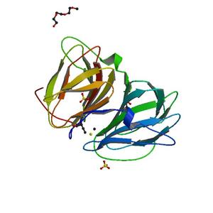
| Title | Crystal structure of uncharacterized conserved protein with double-stranded beta-helix domain (YP_001338853.1) from Klebsiella pneumoniae subsp. pneumoniae MGH 78578 at 1.80 A resolution |
| Authors | Joint Center for Structural Genomics (JCSG) |
| Bioentity | None |
| R / Rfree | 0.15 / 0.20 |
| Unit cell edges [Å] | 46.85 x 46.85 x 94.94 |
| Unit cell angles [°] | 90.0, 90.0, 120.0 |
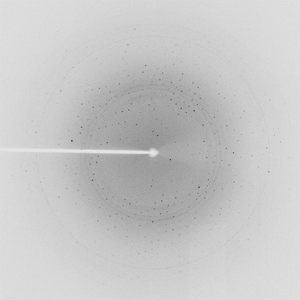
Dataset 93593_1_E1.#### details
| Number of frames | 90 (1 - 90) |
| Distance [mm] | 250.0 |
| Oscillation width [°] | 1.00 |
| Phi [°] | 42.0 |
| Wavelength [Å] | 0.97965 |
| Experiment Date | 2008-10-12 |
| Equipment | 23-ID-B at APS (Advanced Photon Source) |
Dataset 93593_1_E2.#### details
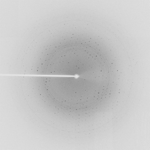
| Number of frames | 90 (1 - 90) |
| Distance [mm] | 250.0 |
| Oscillation width [°] | 1.00 |
| Phi [°] | 42.0 |
| Wavelength [Å] | 0.94645 |
| Experiment Date | 2008-10-12 |
| Equipment | 23-ID-B at APS (Advanced Photon Source) |