Diffraction project datasets MG14579A_3gyd
Project details
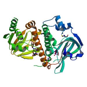
| Title |
Crystal structure of a cyclic nucleotide-binding domain (mfla_1926) from methylobacillus flagellatus kt at 1.79 A resolution |
| Authors |
Joint Center for Structural Genomics (JCSG) |
| Bioentity |
None |
| R / Rfree |
0.17 / 0.21 |
| Unit cell edges [Å] |
60.03 x
39.74 x
70.98
|
| Unit cell angles [°] |
90.0,
101.9,
90.0
|
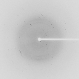
Dataset 105616_1_E1_###.mccd details
| Number of frames |
150 (1 - 150) |
| Distance [mm] |
300.0 |
| Oscillation width [°] |
1.00 |
| Phi [°] |
58.0 |
| Wavelength [Å] |
0.97956 |
| Experiment Date |
2009-01-18 |
| Equipment |
BL9-2
at SSRL (Stanford Synchrotron Radiation Laboratory)
|
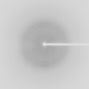
Dataset 105616_1_E2_###.mccd details
| Number of frames |
150 (1 - 150) |
| Distance [mm] |
300.0 |
| Oscillation width [°] |
1.00 |
| Phi [°] |
58.0 |
| Wavelength [Å] |
0.91837 |
| Experiment Date |
2009-01-18 |
| Equipment |
BL9-2
at SSRL (Stanford Synchrotron Radiation Laboratory)
|
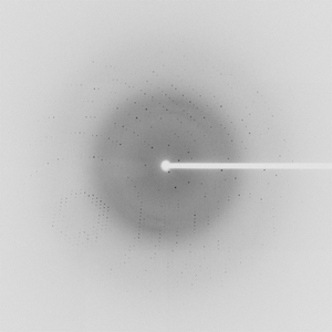
Dataset 105616_2_###.mccd details
| Number of frames |
150 (1 - 150) |
| Distance [mm] |
230.0 |
| Oscillation width [°] |
1.00 |
| Phi [°] |
58.0 |
| Wavelength [Å] |
0.97944 |
| Experiment Date |
2009-01-18 |
| Equipment |
BL9-2
at SSRL (Stanford Synchrotron Radiation Laboratory)
|