6890 results
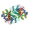
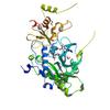
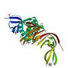
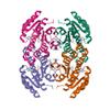
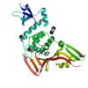
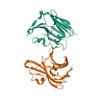
X-ray diffraction data for the Crystal structure of beta-ketoacyl-ACP synthase II (FabF) from Vibrio Cholerae (space group P43) at 2.2 Angstrom
First author:
J. Hou
Resolution: 2.20 Å
R/Rfree: 0.17/0.20
Resolution: 2.20 Å
R/Rfree: 0.17/0.20
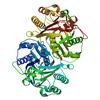
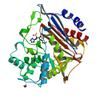
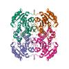
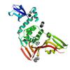
X-ray diffraction data for the Crystal structure of beta-ketoacyl-ACP synthase II (FabF) from Vibrio Cholerae (space group P212121) at 1.75 Angstrom
First author:
J. Hou
Resolution: 1.75 Å
R/Rfree: 0.20/0.23
Resolution: 1.75 Å
R/Rfree: 0.20/0.23
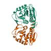
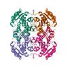
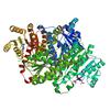
X-ray diffraction data for the Crystal structure of beta-ketoacyl-ACP synthase III (FabH) from Vibrio Cholerae in complex with Coenzyme A

First author:
J. Hou
Resolution: 1.78 Å
R/Rfree: 0.17/0.20
Resolution: 1.78 Å
R/Rfree: 0.17/0.20
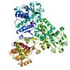
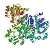
X-ray diffraction data for the Crystal structure of beta-ketoacyl-(acyl carrier protein) synthase III-2 (FabH2) from Vibrio cholerae
First author:
J. Hou
Resolution: 1.88 Å
R/Rfree: 0.17/0.20
Resolution: 1.88 Å
R/Rfree: 0.17/0.20
X-ray diffraction data for the Beta-ketoacyl-(acyl carrier protein) synthase III-2 (FabH2) from Vibrio cholerae soaked with Acetyl-CoA
First author:
J. Hou
Resolution: 2.20 Å
R/Rfree: 0.17/0.20
Resolution: 2.20 Å
R/Rfree: 0.17/0.20
X-ray diffraction data for the Beta-ketoacyl-acyl carrier protein synthase III-2 (FabH2)(C113A) from Vibrio cholerae
First author:
J. Hou
Resolution: 1.61 Å
R/Rfree: 0.15/0.18
Resolution: 1.61 Å
R/Rfree: 0.15/0.18
X-ray diffraction data for the Beta-ketoacyl-ACP synthase III -2 (FabH2) (C113A) from Vibrio Cholerae soaked with octanoyl-CoA: conformational changes without clearly bound substrate
First author:
J. Hou
Resolution: 2.30 Å
R/Rfree: 0.20/0.23
Resolution: 2.30 Å
R/Rfree: 0.20/0.23
X-ray diffraction data for the Crystal structure of C103A mutant of DJ-1 superfamily protein STM1931 from Salmonella typhimurium
First author:
I.A. Shumilin
Gene name: araH
Resolution: 2.20 Å
R/Rfree: 0.17/0.20
Gene name: araH
Resolution: 2.20 Å
R/Rfree: 0.17/0.20
X-ray diffraction data for the Crystal structure of anabolic ornithine carbamoyltransferase from Bacillus anthracis in complex with carbamoyl phosphate and L-norvaline
First author:
I.G. Shabalin
Gene name: argF
Resolution: 1.74 Å
R/Rfree: 0.15/0.17
Gene name: argF
Resolution: 1.74 Å
R/Rfree: 0.15/0.17
X-ray diffraction data for the Crystal Structure of NSP15 Endoribonuclease from SARS CoV-2 in the Complex with potential repurposing drug Tipiracil
First author:
Y. Kim
Resolution: 1.85 Å
R/Rfree: 0.17/0.19
Resolution: 1.85 Å
R/Rfree: 0.17/0.19
X-ray diffraction data for the GBAA_1210 protein, a putative adenylate cyclase, from Bacillus anthracis

First author:
J. Osipiuk
Resolution: 2.14 Å
R/Rfree: 0.20/0.25
Resolution: 2.14 Å
R/Rfree: 0.20/0.25
X-ray diffraction data for the Non-structural protein 10 (nsp10) from SARS CoV-2 in complex with fragment VT00239
First author:
V.O. Talibov
Resolution: 1.95 Å
R/Rfree: 0.17/0.20
Resolution: 1.95 Å
R/Rfree: 0.17/0.20
X-ray diffraction data for the Structure of yncA, a putative ACETYLTRANSFERASE from Salmonella typhimurium
First author:
A.U. Singer
Gene name: argC
Resolution: 1.75 Å
R/Rfree: 0.16/0.20
Gene name: argC
Resolution: 1.75 Å
R/Rfree: 0.16/0.20
X-ray diffraction data for the The crystal structure of Papain-Like Protease of SARS CoV-2 , C111S mutant, at room temperature
First author:
J. Osipiuk
Gene name: orf1ab
Resolution: 2.48 Å
R/Rfree: 0.15/0.19
Gene name: orf1ab
Resolution: 2.48 Å
R/Rfree: 0.15/0.19
X-ray diffraction data for the Class C beta-lactamase from Escherichia coli
First author:
C. Chang
Gene name: None
Resolution: 1.22 Å
R/Rfree: 0.16/0.18
Gene name: None
Resolution: 1.22 Å
R/Rfree: 0.16/0.18
First author:
M.P. Czub
Resolution: 2.40 Å
R/Rfree: 0.20/0.25
Resolution: 2.40 Å
R/Rfree: 0.20/0.25
X-ray diffraction data for the 2.0 Angstrom Resolution Crystal Structure of Nsp16-Nsp10 Heterodimer from SARS-CoV-2 in Complex with S-Adenosyl-L-Homocysteine
First author:
G. Minasov
Gene name: orf1ab
Resolution: 2.00 Å
R/Rfree: 0.17/0.19
Gene name: orf1ab
Resolution: 2.00 Å
R/Rfree: 0.17/0.19
X-ray diffraction data for the 1.98 Angstrom Resolution Crystal Structure of NSP16-NSP10 Heterodimer from SARS-CoV-2 in Complex with Sinefungin
First author:
G. Minasov
Gene name: orf1ab
Resolution: 1.98 Å
R/Rfree: 0.16/0.18
Gene name: orf1ab
Resolution: 1.98 Å
R/Rfree: 0.16/0.18
X-ray diffraction data for the Crystal Structure of Nsp16-Nsp10 Heterodimer from SARS-CoV-2 in Complex with 7-methyl-GpppA and S-adenosyl-L-homocysteine.
First author:
G. Minasov
Gene name: orf1ab
Resolution: 2.10 Å
R/Rfree: 0.17/0.19
Gene name: orf1ab
Resolution: 2.10 Å
R/Rfree: 0.17/0.19
X-ray diffraction data for the Crystal Structure of Nsp16-Nsp10 Heterodimer from SARS-CoV-2 with 7-methyl-GpppA and S-adenosyl-L-homocysteine in the Active Site and Sulfates in the mRNA Binding Groove.
First author:
G. Minasov
Gene name: orf1ab
Resolution: 2.25 Å
R/Rfree: 0.16/0.19
Gene name: orf1ab
Resolution: 2.25 Å
R/Rfree: 0.16/0.19
X-ray diffraction data for the Crystal Structure of Nsp16-Nsp10 from SARS-CoV-2 in Complex with 7-methyl-GpppA and S-Adenosylmethionine.
First author:
G. Minasov
Gene name: orf1ab
Resolution: 2.00 Å
R/Rfree: 0.16/0.18
Gene name: orf1ab
Resolution: 2.00 Å
R/Rfree: 0.16/0.18
X-ray diffraction data for the 2.05 Angstrom Resolution Crystal Structure of C-terminal Dimerization Domain of Nucleocapsid Phosphoprotein from SARS-CoV-2

First author:
G. Minasov
Gene name: N
Resolution: 2.05 Å
R/Rfree: 0.19/0.23
Gene name: N
Resolution: 2.05 Å
R/Rfree: 0.19/0.23
X-ray diffraction data for the Crystal structure of Class D beta-lactamase from Klebsiella quasipneumoniae in complex with avibactam
First author:
C. Chang
Gene name: None
Resolution: 1.85 Å
R/Rfree: 0.18/0.22
Gene name: None
Resolution: 1.85 Å
R/Rfree: 0.18/0.22
X-ray diffraction data for the Crystal structure of uncharacterized protein ECL_02694
First author:
C. Chang
Resolution: 1.45 Å
R/Rfree: 0.15/0.18
Resolution: 1.45 Å
R/Rfree: 0.15/0.18
X-ray diffraction data for the Tryptophan--tRNA ligase from Haemophilus influenzae.
First author:
J. Osipiuk
Gene name: trpS
Resolution: 2.05 Å
R/Rfree: 0.19/0.24
Gene name: trpS
Resolution: 2.05 Å
R/Rfree: 0.19/0.24
X-ray diffraction data for the Beta-lactamase from Escherichia coli str. Sakai
First author:
J. Osipiuk
Gene name: ampC
Resolution: 1.60 Å
R/Rfree: 0.14/0.16
Gene name: ampC
Resolution: 1.60 Å
R/Rfree: 0.14/0.16
X-ray diffraction data for the Crystal structure of ornithine carbamoyltransferase from Salmonella enterica
First author:
C. Chang
Gene name: argI
Resolution: 1.90 Å
R/Rfree: 0.17/0.19
Gene name: argI
Resolution: 1.90 Å
R/Rfree: 0.17/0.19
X-ray diffraction data for the Putative oxidoreductase from Escherichia coli str. K-12
First author:
J. Osipiuk
Gene name: yohF
Resolution: 1.36 Å
R/Rfree: 0.12/0.17
Gene name: yohF
Resolution: 1.36 Å
R/Rfree: 0.12/0.17
X-ray diffraction data for the Succinyl-CoA synthase from Francisella tularensis
First author:
J. Osipiuk
Resolution: 1.76 Å
R/Rfree: 0.19/0.22
Resolution: 1.76 Å
R/Rfree: 0.19/0.22
X-ray diffraction data for the Putative Antitoxin HicB3 from Escherichia coli str. K-12 substr. DH10B
First author:
J. Osipiuk
Gene name: ydcQ
Resolution: 1.90 Å
R/Rfree: 0.20/0.25
Gene name: ydcQ
Resolution: 1.90 Å
R/Rfree: 0.20/0.25
X-ray diffraction data for the Crystal structure of Equine Serum Albumin complex with ketoprofen
First author:
M.P. Czub
Gene name: ALB
Resolution: 2.45 Å
R/Rfree: 0.18/0.23
Gene name: ALB
Resolution: 2.45 Å
R/Rfree: 0.18/0.23
X-ray diffraction data for the Crystal structure of Equine Serum Albumin complex with ibuprofen
First author:
M.P. Czub
Gene name: ALB
Resolution: 2.25 Å
R/Rfree: 0.19/0.24
Gene name: ALB
Resolution: 2.25 Å
R/Rfree: 0.19/0.24
X-ray diffraction data for the Crystal structure of Equine Serum Albumin complex with 6-MNA
First author:
M.P. Czub
Gene name: ALB
Resolution: 2.65 Å
R/Rfree: 0.19/0.26
Gene name: ALB
Resolution: 2.65 Å
R/Rfree: 0.19/0.26
X-ray diffraction data for the OmpA-like domain of FopA1 from Francisella tularensis subsp. tularensis SCHU S4
First author:
K. Michalska
Gene name: fopA1
Resolution: 2.36 Å
R/Rfree: 0.21/0.24
Gene name: fopA1
Resolution: 2.36 Å
R/Rfree: 0.21/0.24
X-ray diffraction data for the Crystal structure of 3-deoxy-D-arabinoheptulosonate-7-phosphate synthase/phospho-2-dehydro-3-deoxyheptonate aldolase (Aro3) from Candida auris
First author:
K. Michalska
Resolution: 2.49 Å
R/Rfree: 0.18/0.23
Resolution: 2.49 Å
R/Rfree: 0.18/0.23
X-ray diffraction data for the Crystal structure of acetoin dehydrogenase from Enterobacter cloacae
First author:
C. Chang
Resolution: 2.20 Å
R/Rfree: 0.18/0.24
Resolution: 2.20 Å
R/Rfree: 0.18/0.24
X-ray diffraction data for the Crystal structure of unknown function protein yfdX from Shigella flexneri
First author:
C. Chang
Resolution: 1.80 Å
R/Rfree: 0.17/0.20
Resolution: 1.80 Å
R/Rfree: 0.17/0.20
X-ray diffraction data for the Crystal structure of RNA binding domain of nucleocapsid phosphoprotein from SARS coronavirus 2
First author:
C. Chang
Gene name: N
Resolution: 1.70 Å
R/Rfree: 0.16/0.21
Gene name: N
Resolution: 1.70 Å
R/Rfree: 0.16/0.21
X-ray diffraction data for the Crystal Structure of Motility Associated Killing Factor B from Vibrio cholerae
First author:
Y. Kim
Gene name: makB
Resolution: 2.58 Å
R/Rfree: 0.28/0.29
Gene name: makB
Resolution: 2.58 Å
R/Rfree: 0.28/0.29
X-ray diffraction data for the The crystal structure of papain-like protease of SARS CoV-2
First author:
J. Osipiuk
Gene name: orf1ab
Resolution: 2.70 Å
R/Rfree: 0.24/0.28
Gene name: orf1ab
Resolution: 2.70 Å
R/Rfree: 0.24/0.28
X-ray diffraction data for the 1.80 Angstrom Resolution Crystal Structure of NSP16 - NSP10 Complex from SARS-CoV-2
First author:
G. Minasov
Gene name: orf1ab
Resolution: 1.80 Å
R/Rfree: 0.15/0.16
Gene name: orf1ab
Resolution: 1.80 Å
R/Rfree: 0.15/0.16
X-ray diffraction data for the 1.95 Angstrom Resolution Crystal Structure of NSP10 - NSP16 Complex from SARS-CoV-2
First author:
G. Minasov
Gene name: orf1ab
Resolution: 1.95 Å
R/Rfree: 0.16/0.17
Gene name: orf1ab
Resolution: 1.95 Å
R/Rfree: 0.16/0.17
X-ray diffraction data for the Crystal Structure of NSP15 Endoribonuclease from SARS CoV-2.
First author:
Y. Kim
Gene name: orf1ab
Resolution: 2.20 Å
R/Rfree: 0.16/0.18
Gene name: orf1ab
Resolution: 2.20 Å
R/Rfree: 0.16/0.18
X-ray diffraction data for the Crystal Structure of ADP ribose phosphatase of NSP3 from SARS CoV-2
First author:
Y. Kim
Gene name: orf1ab
Resolution: 2.03 Å
R/Rfree: 0.19/0.23
Gene name: orf1ab
Resolution: 2.03 Å
R/Rfree: 0.19/0.23
X-ray diffraction data for the The 1.9 A Crystal Structure of NSP15 Endoribonuclease from SARS CoV-2 in the Complex with a Citrate
First author:
Y. Kim
Gene name: orf1ab
Resolution: 1.90 Å
R/Rfree: 0.16/0.18
Gene name: orf1ab
Resolution: 1.90 Å
R/Rfree: 0.16/0.18
X-ray diffraction data for the The crystal structure of Nsp9 RNA binding protein of SARS CoV-2
First author:
K. Tan
Gene name: orf1ab
Resolution: 2.95 Å
R/Rfree: 0.24/0.28
Gene name: orf1ab
Resolution: 2.95 Å
R/Rfree: 0.24/0.28
X-ray diffraction data for the Crystal Structure of ADP ribose phosphatase of NSP3 from SARS CoV-2 in the complex with ADP ribose
First author:
K. Michalska
Gene name: orf1ab
Resolution: 1.50 Å
R/Rfree: 0.15/0.17
Gene name: orf1ab
Resolution: 1.50 Å
R/Rfree: 0.15/0.17
X-ray diffraction data for the Crystal Structure of the methyltransferase-stimulatory factor complex of NSP16 and NSP10 from SARS CoV-2.
First author:
Y. Kim
Resolution: 2.00 Å
R/Rfree: 0.17/0.19
Resolution: 2.00 Å
R/Rfree: 0.17/0.19
X-ray diffraction data for the CRYSTAL STRUCTURE OF CLASS C BETA-LACTAMASE FROM ESCHERICHIA COLI IN COMPLEX WITH TAZOBACTAM
First author:
N.MALTSEVA C.CHANG
Gene name: None
Resolution: 2.70 Å
R/Rfree: 0.19/0.25
Gene name: None
Resolution: 2.70 Å
R/Rfree: 0.19/0.25
X-ray diffraction data for the Crystal Structure of ADP ribose phosphatase of NSP3 from SARS-CoV-2 in the apo form
First author:
K. Michalska
Gene name: orf1ab
Resolution: 1.35 Å
R/Rfree: 0.10/0.14
Gene name: orf1ab
Resolution: 1.35 Å
R/Rfree: 0.10/0.14
X-ray diffraction data for the Crystal Structure of ADP ribose phosphatase of NSP3 from SARS-CoV-2 in complex with MES
First author:
K. Michalska
Gene name: orf1ab
Resolution: 1.07 Å
R/Rfree: 0.13/0.15
Gene name: orf1ab
Resolution: 1.07 Å
R/Rfree: 0.13/0.15
X-ray diffraction data for the class C beta-lactamase from Escherichia coli in complex with cephalothin
First author:
C. Chang
Gene name: None
Resolution: 1.40 Å
R/Rfree: 0.16/0.18
Gene name: None
Resolution: 1.40 Å
R/Rfree: 0.16/0.18
X-ray diffraction data for the Class C beta-lactamase from Acinetobacter baumannii in complex with 4-(Ethyl(methyl)carbamoyl)phenyl boronic acid

First author:
C. Chang
Gene name: None
Resolution: 2.15 Å
R/Rfree: 0.19/0.24
Gene name: None
Resolution: 2.15 Å
R/Rfree: 0.19/0.24
X-ray diffraction data for the Class A beta-lactamase from Micromonospora aurantiaca ATCC 27029
First author:
C. Chang
Gene name: None
Resolution: 1.40 Å
R/Rfree: 0.14/0.16
Gene name: None
Resolution: 1.40 Å
R/Rfree: 0.14/0.16
X-ray diffraction data for the Crystal structure of RNA-binding domain of nucleocapsid phosphoprotein from SARS CoV-2, monoclinic crystal form
First author:
C. Chang
Gene name: N
Resolution: 2.67 Å
R/Rfree: 0.20/0.25
Gene name: N
Resolution: 2.67 Å
R/Rfree: 0.20/0.25
X-ray diffraction data for the Crystal structure of Human Serum Albumin complex with JMS-053
First author:
M.P. Czub
Gene name: ALB
Resolution: 2.20 Å
R/Rfree: 0.19/0.25
Gene name: ALB
Resolution: 2.20 Å
R/Rfree: 0.19/0.25
X-ray diffraction data for the The crystal structure of Papain-Like Protease of SARS CoV-2 , C111S mutant
First author:
J. Osipiuk
Gene name: orf1ab
Resolution: 1.60 Å
R/Rfree: 0.12/0.16
Gene name: orf1ab
Resolution: 1.60 Å
R/Rfree: 0.12/0.16
X-ray diffraction data for the The crystal structure of Papain-Like Protease of SARS CoV-2 , P3221 space group
First author:
J. Osipiuk
Gene name: orf1ab
Resolution: 1.79 Å
R/Rfree: 0.16/0.17
Gene name: orf1ab
Resolution: 1.79 Å
R/Rfree: 0.16/0.17
X-ray diffraction data for the Crystal Structure of NSP15 Endoribonuclease from SARS CoV-2 in the Complex with 3'-uridinemonophosphate

First author:
C. Chang
Resolution: 1.85 Å
R/Rfree: 0.17/0.19
Resolution: 1.85 Å
R/Rfree: 0.17/0.19
X-ray diffraction data for the Crystal structure of 3-ketoacyl-(acyl-carrier-protein) reductase (FabG)(Y155F) from Vibrio cholerae
First author:
J. Hou
Gene name: fabG
Resolution: 2.45 Å
R/Rfree: 0.19/0.23
Gene name: fabG
Resolution: 2.45 Å
R/Rfree: 0.19/0.23
X-ray diffraction data for the Structure of beta-ketoacyl-ACP synthase I (FabB) from Vibrio Cholerae
First author:
J. Hou
Gene name: fabB
Resolution: 2.01 Å
R/Rfree: 0.15/0.18
Gene name: fabB
Resolution: 2.01 Å
R/Rfree: 0.15/0.18
X-ray diffraction data for the Crystal structure of beta-ketoacyl-acyl carrier protein reductase (FabG)(G141A) from Vibrio cholerae
First author:
J. Hou
Gene name: fabG
Resolution: 2.40 Å
R/Rfree: 0.20/0.24
Gene name: fabG
Resolution: 2.40 Å
R/Rfree: 0.20/0.24
X-ray diffraction data for the Crystal structure of anabolic ornithine carbamoyltransferase from Vibrio vulnificus in complex with citrulline and inorganic phosphate
First author:
I.G. Shabalin
Resolution: 2.08 Å
R/Rfree: 0.16/0.19
Resolution: 2.08 Å
R/Rfree: 0.16/0.19
X-ray diffraction data for the Crystal structure of 3-oxoacyl-[acyl-carrier protein]reductase (FabG)from Listeria monocytogenes in complex with NADP+
First author:
J. Hou
Gene name: fabG
Resolution: 1.92 Å
R/Rfree: 0.16/0.20
Gene name: fabG
Resolution: 1.92 Å
R/Rfree: 0.16/0.20
X-ray diffraction data for the Crystal structure of anabolic ornithine carbamoyltransferase from Vibrio vulnificus in complex with carbamoyl phosphate and L-norvaline
First author:
I.G. Shabalin
Resolution: 1.70 Å
R/Rfree: 0.14/0.17
Resolution: 1.70 Å
R/Rfree: 0.14/0.17
X-ray diffraction data for the Crystal structure of transcriptional regulator VanUg, Form II
First author:
P.J. Stogios
Gene name: vanUG
Resolution: 1.12 Å
R/Rfree: 0.14/0.16
Gene name: vanUG
Resolution: 1.12 Å
R/Rfree: 0.14/0.16
X-ray diffraction data for the Crystal structure of unliganded anabolic ornithine carbamoyltransferase from Vibrio vulnificus at 1.86 A resolution
First author:
I.G. Shabalin
Resolution: 1.86 Å
R/Rfree: 0.16/0.20
Resolution: 1.86 Å
R/Rfree: 0.16/0.20
X-ray diffraction data for the Crystal structure of disulfide bond oxidoreductase DsbA1 from Legionella pneumophila
First author:
I.A. Shumilin
Gene name: dsbA
Resolution: 1.88 Å
R/Rfree: 0.16/0.20
Gene name: dsbA
Resolution: 1.88 Å
R/Rfree: 0.16/0.20
X-ray diffraction data for the Crystal structure of a putative 3-oxoacyl-[acyl-carrier protein]reductase from Helicobacter pylori 26695
First author:
J. Hou
Resolution: 2.54 Å
R/Rfree: 0.23/0.27
Resolution: 2.54 Å
R/Rfree: 0.23/0.27
X-ray diffraction data for the Crystal structure of a putative diacylglycerol kinase from Bacillus anthracis str. Sterne
First author:
J. Hou
Gene name: -
Resolution: 2.50 Å
R/Rfree: 0.21/0.24
Gene name: -
Resolution: 2.50 Å
R/Rfree: 0.21/0.24
X-ray diffraction data for the Crystal structure of transcriptional regulator VanUg, Form I
First author:
P.J. Stogios
Gene name: vanUG
Resolution: 1.70 Å
R/Rfree: 0.16/0.20
Gene name: vanUG
Resolution: 1.70 Å
R/Rfree: 0.16/0.20
X-ray diffraction data for the Crystal structure of Tryptophanyl-tRNA synthetase from Vibrio cholerae with an endogenous tryptophan
First author:
D.R. Cooper
Resolution: 1.50 Å
R/Rfree: 0.16/0.18
Resolution: 1.50 Å
R/Rfree: 0.16/0.18
X-ray diffraction data for the Crystal structure of beta-ketoacyl-acyl carrier protein reductase (FabG) from Vibrio cholerae in complex with NADPH
First author:
J. Hou
Gene name: fabG
Resolution: 2.06 Å
R/Rfree: 0.15/0.17
Gene name: fabG
Resolution: 2.06 Å
R/Rfree: 0.15/0.17
X-ray diffraction data for the Crystal structure of a putative 3-oxoacyl-[acyl-carrier protein]reductase from Escherichia coli strain CFT073 complexed with NADP+ at 2.5 A resolution
First author:
J. Hou
Resolution: 2.50 Å
R/Rfree: 0.18/0.22
Resolution: 2.50 Å
R/Rfree: 0.18/0.22
X-ray diffraction data for the Crystal structure of a putative 3-oxoacyl-[acyl-carrier protein]reductase from Escherichia coli strain CFT073 complexed with NADP+ at 2.1 A resolution
First author:
J. Hou
Resolution: 2.10 Å
R/Rfree: 0.17/0.19
Resolution: 2.10 Å
R/Rfree: 0.17/0.19
X-ray diffraction data for the Structure of 3-ketoacyl-(acyl-carrier-protein)reductase (FabG) from Vibrio cholerae O1 complexed with NADP+ (space group P62)
First author:
J. Hou
Gene name: fabG
Resolution: 1.95 Å
R/Rfree: 0.13/0.16
Gene name: fabG
Resolution: 1.95 Å
R/Rfree: 0.13/0.16
X-ray diffraction data for the Crystal structure of anabolic ornithine carbamoyltransferase from Campylobacter jejuni subsp. jejuni NCTC 11168
First author:
I.G. Shabalin
Gene name: argF
Resolution: 2.70 Å
R/Rfree: 0.20/0.24
Gene name: argF
Resolution: 2.70 Å
R/Rfree: 0.20/0.24
X-ray diffraction data for the Crystal structure of 3-ketoacyl-(acyl-carrier-protein) reductase (FabG)(G92D) from Vibrio cholerae
First author:
J. Hou
Gene name: fabG
Resolution: 1.75 Å
R/Rfree: 0.16/0.19
Gene name: fabG
Resolution: 1.75 Å
R/Rfree: 0.16/0.19
X-ray diffraction data for the Crystal structure of Phosphoglycerate Kinase from Campylobacter jejuni.
First author:
E.V. Filippova
Gene name: pgk
Resolution: 2.15 Å
R/Rfree: 0.19/0.24
Gene name: pgk
Resolution: 2.15 Å
R/Rfree: 0.19/0.24
X-ray diffraction data for the Crystal structure of anabolic ornithine carbamoyltransferase from Vibrio vulnificus in complex with carbamoyl phosphate
First author:
I.G. Shabalin
Resolution: 2.17 Å
R/Rfree: 0.17/0.21
Resolution: 2.17 Å
R/Rfree: 0.17/0.21
X-ray diffraction data for the Crystal structure of putative 3-ketoacyl-(acyl-carrier-protein) reductase from Vibrio cholerae O1 biovar eltor str. N16961 in complex with NADP+
First author:
J. Hou
Gene name: fabG
Resolution: 1.60 Å
R/Rfree: 0.15/0.18
Gene name: fabG
Resolution: 1.60 Å
R/Rfree: 0.15/0.18
X-ray diffraction data for the Crystal structure of a putative 3-oxoacyl-[acyl-carrier protein]reductase from Helicobacter pylori 26695 complexed with NAD+
First author:
J. Hou
Resolution: 2.40 Å
R/Rfree: 0.22/0.25
Resolution: 2.40 Å
R/Rfree: 0.22/0.25
X-ray diffraction data for the Crystal structure of 3-ketoacyl-(acyl-carrier-protein) reductase (FabG) from Vibrio cholerae
First author:
J. Hou
Gene name: fabG
Resolution: 2.00 Å
R/Rfree: 0.14/0.18
Gene name: fabG
Resolution: 2.00 Å
R/Rfree: 0.14/0.18
X-ray diffraction data for the Crystal structure of 3-ketoacyl-(acyl-carrier-protein) reductase (FabG)(G92A) from Vibrio cholerae
First author:
J. Hou
Gene name: fabG
Resolution: 1.80 Å
R/Rfree: 0.16/0.21
Gene name: fabG
Resolution: 1.80 Å
R/Rfree: 0.16/0.21
X-ray diffraction data for the Crystal structure of 3-ketoacyl-(acyl-carrier-protein) reductase (FabG)(F187A) from Vibrio cholerae

First author:
J. Hou
Gene name: fabG
Resolution: 2.10 Å
R/Rfree: 0.16/0.19
Gene name: fabG
Resolution: 2.10 Å
R/Rfree: 0.16/0.19
X-ray diffraction data for the Crystal structure of anabolic ornithine carbamoyltransferase from Vibrio vulnificus in complex with carbamoylphosphate and arginine
First author:
I.G. Shabalin
Resolution: 1.85 Å
R/Rfree: 0.15/0.19
Resolution: 1.85 Å
R/Rfree: 0.15/0.19
X-ray diffraction data for the A putative diacylglycerol kinase from Bacillus anthracis str. Sterne
First author:
J. Hou
Gene name: -
Resolution: 2.16 Å
R/Rfree: 0.20/0.23
Gene name: -
Resolution: 2.16 Å
R/Rfree: 0.20/0.23
X-ray diffraction data for the The crystal structure of Papain-Like Protease of SARS CoV-2 , C111S mutant, in complex with PLP_Snyder457 inhibitor
First author:
J. Osipiuk
Resolution: 2.09 Å
R/Rfree: 0.19/0.20
Resolution: 2.09 Å
R/Rfree: 0.19/0.20
X-ray diffraction data for the The crystal structure of Papain-Like Protease of SARS CoV-2 , C111S mutant, in complex with PLP_Snyder495 inhibitor
First author:
J. Osipiuk
Resolution: 1.95 Å
R/Rfree: 0.17/0.19
Resolution: 1.95 Å
R/Rfree: 0.17/0.19
X-ray diffraction data for the The crystal structure of Papain-Like Protease of SARS CoV-2 , C111S mutant, in complex with PLP_Snyder530 inhibitor
First author:
J. Osipiuk
Resolution: 2.05 Å
R/Rfree: 0.19/0.20
Resolution: 2.05 Å
R/Rfree: 0.19/0.20
X-ray diffraction data for the The crystal structure of Papain-Like Protease of SARS CoV-2 in complex with PLP_Snyder530 inhibitor
First author:
J. Osipiuk
Resolution: 2.30 Å
R/Rfree: 0.21/0.24
Resolution: 2.30 Å
R/Rfree: 0.21/0.24
X-ray diffraction data for the The crystal structure of Papain-Like Protease of SARS CoV-2 in complex with PLP_Snyder441 inhibitor
First author:
J. Osipiuk
Resolution: 1.93 Å
R/Rfree: 0.19/0.21
Resolution: 1.93 Å
R/Rfree: 0.19/0.21
X-ray diffraction data for the Crystal structure of 3-ketoacyl-(acyl-carrier-protein) reductase (FabG) (G141A) from Vibrio cholerae in complex with NADPH
First author:
J. Hou
Gene name: fabG
Resolution: 2.21 Å
R/Rfree: 0.21/0.24
Gene name: fabG
Resolution: 2.21 Å
R/Rfree: 0.21/0.24
X-ray diffraction data for the Structure of a putative lipoprotein from Staphylococcus aureus subsp. aureus NCTC 8325
First author:
E.V. Filippova
Resolution: 3.08 Å
R/Rfree: 0.23/0.28
Resolution: 3.08 Å
R/Rfree: 0.23/0.28
X-ray diffraction data for the Crystal structure of Human Serum Albumin in complex with ketoprofen
First author:
M.P. Czub
Gene name: ALB
Resolution: 2.60 Å
R/Rfree: 0.18/0.23
Gene name: ALB
Resolution: 2.60 Å
R/Rfree: 0.18/0.23
X-ray diffraction data for the 1.76 Angstrom Crystal Structure of GTP-binding Protein Der from Coxiella burnetii in Complex with GDP.
First author:
G. Minasov
Resolution: 1.76 Å
R/Rfree: 0.17/0.20
Resolution: 1.76 Å
R/Rfree: 0.17/0.20
X-ray diffraction data for the 2.35 Angstrom resolution structure of WecB (VC0917), a UDP-N-acetylglucosamine 2-epimerase from Vibrio cholerae.
First author:
G. Minasov
Gene name: wecB
Resolution: 2.35 Å
R/Rfree: 0.17/0.23
Gene name: wecB
Resolution: 2.35 Å
R/Rfree: 0.17/0.23
X-ray diffraction data for the Crystal structure of chaperone CsaA form Bacillus anthracis str. Ames
First author:
B. Nocek
Gene name: csaA
Resolution: 1.50 Å
R/Rfree: 0.17/0.20
Gene name: csaA
Resolution: 1.50 Å
R/Rfree: 0.17/0.20
X-ray diffraction data for the The Crystal Structure of a Dihydroorotase from Staphylococcus aureus
First author:
J.S. Brunzelle
Gene name: pyrC
Resolution: 2.00 Å
R/Rfree: 0.19/0.24
Gene name: pyrC
Resolution: 2.00 Å
R/Rfree: 0.19/0.24
X-ray diffraction data for the Inosine 5'-monophosphate dehydrogenase from Vibrio cholerae, deletion mutant, complexed with IMP.
First author:
N.Maltseva 'J.Osipiuk
Gene name: guaB
Resolution: 2.15 Å
R/Rfree: 0.21/0.26
Gene name: guaB
Resolution: 2.15 Å
R/Rfree: 0.21/0.26