Diffraction project datasets IDP01750_3s19
- Method: MIR
- Resolution: 1.5009 Å
- Space group: P 1
PDB website for 3S19
PDB validation report for 3S19
doi:10.18430/m33s19
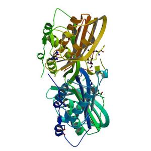
Project details
| Title | Crystal Structure of the R262L mutant of 7-cyano-7-deazaguanine reductase, QueF from Vibrio cholerae complexed with preQ0 |
| Authors | Kim, Y., Zhou, M., Gu, M., Anderson, W.F., Joachimiak, A., Center for Structural Genomics of Infectious Diseases (CSGID) |
| R / Rfree | 0.15 / 0.18 |
| Unit cell edges [Å] | 71.33 x 71.33 x 71.33 |
| Unit cell angles [°] | 110.0, 119.5, 99.5 |
Dataset 262l-q0-m1d7g-peak.####.img details
| Number of frames | 222 (1 - 222) |
| Distance [mm] | 220.0 |
| Oscillation width [°] | 1.00 |
| Omega [°] | -120.0 |
| Kappa / Chi [°] | 0.0003 |
| Phi [°] | -0.0037 |
| Wavelength [Å] | 0.97929 |
| Experiment Date | 2010-12-10 |
| Equipment | 19-ID at APS (Advanced Photon Source) |