417 results
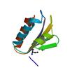
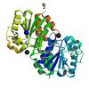
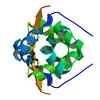
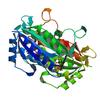
X-ray diffraction data for the Crystal Structure of a Phosphocarrier Protein HPr from Bacillus anthracis str. Ames
First author:
J.S. Brunzelle
Gene name: ptsH-2
Resolution: 1.15 Å
R/Rfree: 0.16/0.18
Gene name: ptsH-2
Resolution: 1.15 Å
R/Rfree: 0.16/0.18
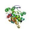
X-ray diffraction data for the 0.95A Resolution Structure of a Histidine Triad Protein from Clostridium difficile
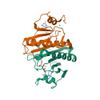
First author:
S.M. Anderson
Resolution: 0.95 Å
R/Rfree: 0.14/0.15
Resolution: 0.95 Å
R/Rfree: 0.14/0.15
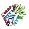
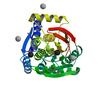
X-ray diffraction data for the Structure of phosphotransferase enzyme II, A component from Yersinia pestis CO92 at 1.2 A resolution

First author:
E.V. Filippova
Resolution: 1.20 Å
R/Rfree: 0.15/0.18
Resolution: 1.20 Å
R/Rfree: 0.15/0.18
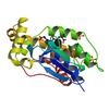
X-ray diffraction data for the 1.05 Angstrom crystal structure of an amino acid ABC transporter substrate-binding protein AbpA from Streptococcus pneumoniae Canada MDR_19A bound to L-arginine
First author:
P.J. Stogios
Resolution: 1.05 Å
R/Rfree: 0.17/0.18
Resolution: 1.05 Å
R/Rfree: 0.17/0.18
X-ray diffraction data for the 1.2 Angstrom Crystal Structure of the Glutaredoxin 2 (grxB) from Salmonella typhimurium in complex with Glutathione
First author:
G. Minasov
Gene name: grxB
Resolution: 1.20 Å
R/Rfree: 0.12/0.14
Gene name: grxB
Resolution: 1.20 Å
R/Rfree: 0.12/0.14
X-ray diffraction data for the Crystal structure of PriA from Actinomyces urogenitalis
First author:
K. MICHALSKA
Resolution: 1.05 Å
R/Rfree: 0.12/0.14
Resolution: 1.05 Å
R/Rfree: 0.12/0.14
X-ray diffraction data for the 1.18 Angstrom resolution crystal structure of uncharacterized protein lmo1340 from Listeria monocytogenes EGD-e
First author:
A.S. Halavaty
Gene name: -
Resolution: 1.18 Å
R/Rfree: 0.15/0.18
Gene name: -
Resolution: 1.18 Å
R/Rfree: 0.15/0.18
X-ray diffraction data for the 1.1 Angstrom Crystal Structure of Hypothetical Protein BA_2335 from Bacillus anthracis
First author:
G. Minasov
Resolution: 1.10 Å
R/Rfree: 0.15/0.18
Resolution: 1.10 Å
R/Rfree: 0.15/0.18
X-ray diffraction data for the Crystal structure of VC2308 protein
First author:
E. Niedzialkowska
Resolution: 1.16 Å
R/Rfree: 0.14/0.17
Resolution: 1.16 Å
R/Rfree: 0.14/0.17
X-ray diffraction data for the Crystal structure of a putative extracellular heme-binding protein (DESPIG_02683) from Desulfovibrio piger ATCC 29098 at 1.24 A resolution
First author:
JOINT CENTER FOR STRUCTURAL GENOMICS (JCSG)
Resolution: 1.24 Å
R/Rfree: 0.13/0.15
Resolution: 1.24 Å
R/Rfree: 0.13/0.15
X-ray diffraction data for the Crystal structure of SusD superfamily protein (YP_001299712.1) from Bacteroides vulgatus ATCC 8482 at 1.13 A resolution
First author:
Joint Center for Structural Genomics (JCSG)
Resolution: 1.13 Å
R/Rfree: 0.12/0.14
Resolution: 1.13 Å
R/Rfree: 0.12/0.14
X-ray diffraction data for the PKD domain of M14-like peptidase from Thermoplasmatales archaeon SCGC AB-540-F20
First author:
K. Michalska
Resolution: 1.23 Å
R/Rfree: 0.14/0.17
Resolution: 1.23 Å
R/Rfree: 0.14/0.17
X-ray diffraction data for the Structure of the YrdA ferripyochelin binding protein from Salmonella enterica
First author:
S.M. Anderson
Gene name: yrdA
Resolution: 1.20 Å
R/Rfree: 0.14/0.16
Gene name: yrdA
Resolution: 1.20 Å
R/Rfree: 0.14/0.16
X-ray diffraction data for the New Delhi Metallo-beta-Lactamase-1 1.05 A structure Complexed with Hydrolyzed Ampicillin
First author:
Y. Kim
Resolution: 1.05 Å
R/Rfree: 0.13/0.16
Resolution: 1.05 Å
R/Rfree: 0.13/0.16
X-ray diffraction data for the Crystal structure of hypothetical protein with ketosteroid isomerase-like protein fold from Catenulispora acidiphila DSM 44928
First author:
E.V. Filippova
Resolution: 1.15 Å
R/Rfree: 0.13/0.14
Resolution: 1.15 Å
R/Rfree: 0.13/0.14
X-ray diffraction data for the Crystal structure of TrpF from Jonesia denitrificans
First author:
K. Michalska
Resolution: 1.09 Å
R/Rfree: 0.13/0.14
Resolution: 1.09 Å
R/Rfree: 0.13/0.14
X-ray diffraction data for the Fatty acid ABC transporter substrate-binding protein from Thermomonospora curvata
First author:
J. Osipiuk
Resolution: 1.15 Å
R/Rfree: 0.12/0.15
Resolution: 1.15 Å
R/Rfree: 0.12/0.15
X-ray diffraction data for the 1.03 Angstrom Crystal Structure of Q236A Mutant Type I Dehydroquinate Dehydratase (aroD) from Salmonella typhimurium
First author:
S.H. Light
Gene name: aroD
Resolution: 1.03 Å
R/Rfree: 0.14/0.16
Gene name: aroD
Resolution: 1.03 Å
R/Rfree: 0.14/0.16
X-ray diffraction data for the Crystal Structure of ThiJ/PfpI Domain Protein from Brachyspira murdochii
First author:
Y. Kim
Resolution: 1.12 Å
R/Rfree: 0.12/0.13
Resolution: 1.12 Å
R/Rfree: 0.12/0.13
X-ray diffraction data for the Crystal structure of a putative ornithine aminotransferase from Toxoplasma gondii ME49 in complex with pyrodoxal-5'-phosphate
First author:
E.V. Filippova
Resolution: 1.20 Å
R/Rfree: 0.13/0.16
Resolution: 1.20 Å
R/Rfree: 0.13/0.16
X-ray diffraction data for the Crystal structure of transcriptional regulator VanUg, Form II
First author:
P.J. Stogios
Gene name: vanUG
Resolution: 1.12 Å
R/Rfree: 0.14/0.16
Gene name: vanUG
Resolution: 1.12 Å
R/Rfree: 0.14/0.16
X-ray diffraction data for the 1.0 Angstrom resolution crystal structure of the branched-chain amino acid transporter substrate binding protein LivJ from Streptococcus pneumoniae str. Canada MDR_19A in complex with Isoleucine
First author:
A.S. Halavaty
Resolution: 1.00 Å
R/Rfree: 0.13/0.15
Resolution: 1.00 Å
R/Rfree: 0.13/0.15
X-ray diffraction data for the Crystal structure of iron uptake ABC transporter substrate-binding protein PiuA from Streptococcus pneumoniae Canada MDR_19A
First author:
P.J. Stogios
Resolution: 1.13 Å
R/Rfree: 0.14/0.17
Resolution: 1.13 Å
R/Rfree: 0.14/0.17
X-ray diffraction data for the 1.02 Angstrom resolution crystal structure of 3-phosphoshikimate 1-carboxyvinyltransferase from Vibrio cholerae in complex with shikimate-3-phosphate (partially photolyzed) and glyphosate
First author:
G. Minasov
Resolution: 1.02 Å
R/Rfree: 0.11/0.14
Resolution: 1.02 Å
R/Rfree: 0.11/0.14
X-ray diffraction data for the 1.1 Angstrom Crystal Structure of Putative Modulator of Drug Activity (MdaB) from Yersinia pestis CO92.
First author:
G. Minasov
Gene name: mdaB
Resolution: 1.10 Å
R/Rfree: 0.10/0.12
Gene name: mdaB
Resolution: 1.10 Å
R/Rfree: 0.10/0.12