Diffraction project datasets WS20613E_3s6e
Project details
| Title |
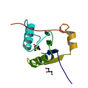
Crystal structure of a RNA binding motif protein 39 (RBM39) from Mus musculuS at 0.95 A resolution |
| Authors |
Joint Center for Structural Genomics (JCSG), Partnership for T-Cell Biology (TCELL) |
| Bioentity |
None |
| R / Rfree |
0.12 / 0.13 |
| Unit cell edges [Å] |
44.70 x
55.14 x
84.60
|
| Unit cell angles [°] |
90.0,
90.0,
90.0
|
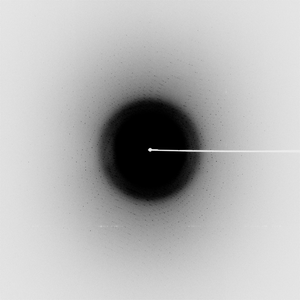
Dataset 165231_1_####.mccd details
| Number of frames |
300 (1 - 300) |
| Distance [mm] |
100.0 |
| Oscillation width [°] |
0.50 |
| Phi [°] |
286.0 |
| Wavelength [Å] |
0.97915 |
| Experiment Date |
2011-03-05 |
| Equipment |
BL14-1
at SSRL (Stanford Synchrotron Radiation Laboratory)
|
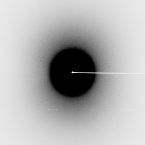
Dataset 165231_2_####.mccd details
| Number of frames |
240 (1 - 240) |
| Distance [mm] |
96.0 |
| Oscillation width [°] |
0.50 |
| Phi [°] |
286.0 |
| Wavelength [Å] |
0.95369 |
| Experiment Date |
2011-03-05 |
| Equipment |
BL14-1
at SSRL (Stanford Synchrotron Radiation Laboratory)
|
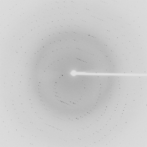
Dataset 165231_3_####.mccd details
| Number of frames |
180 (1 - 180) |
| Distance [mm] |
310.0 |
| Oscillation width [°] |
0.50 |
| Phi [°] |
286.0 |
| Wavelength [Å] |
0.95369 |
| Experiment Date |
2011-03-05 |
| Equipment |
BL14-1
at SSRL (Stanford Synchrotron Radiation Laboratory)
|