402 results
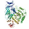
X-ray diffraction data for the Crystal structure of the yhdH oxidoreductase from Salmonella enterica in complex with NADP
First author:
S.M. Anderson
Gene name: yhdH
Resolution: 1.90 Å
R/Rfree: 0.15/0.19
Gene name: yhdH
Resolution: 1.90 Å
R/Rfree: 0.15/0.19
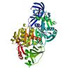
X-ray diffraction data for the Crystal Structure of a propionate kinase from Francisella tularensis subsp. tularensis SCHU S4

First author:
J.S. Brunzelle
Gene name: tdcD
Resolution: 1.98 Å
R/Rfree: 0.16/0.19
Gene name: tdcD
Resolution: 1.98 Å
R/Rfree: 0.16/0.19
X-ray diffraction data for the The crystal structure of kanamycin B dioxygenase (KanJ) from Streptomyces kanamyceticus in complex with nickel, sulfate, soaked with iodide
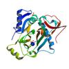
First author:
B. Mrugala
Resolution: 2.10 Å
R/Rfree: 0.17/0.20
Resolution: 2.10 Å
R/Rfree: 0.17/0.20
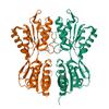
X-ray diffraction data for the Crystal Structure of the Sugar Binding Domain of LacI Family Protein from Klebsiella pneumoniae

X-ray diffraction data for the Structure of a putative aminopeptidase P from Bacillus anthracis
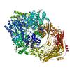
First author:
S.M. Anderson
Resolution: 2.89 Å
R/Rfree: 0.20/0.23
Resolution: 2.89 Å
R/Rfree: 0.20/0.23
X-ray diffraction data for the Crystal structure of the Bacillus anthracis acetyl-CoA acetyltransferase
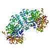
First author:
S.M. Anderson
Resolution: 1.70 Å
R/Rfree: 0.15/0.17
Resolution: 1.70 Å
R/Rfree: 0.15/0.17
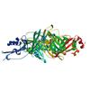
X-ray diffraction data for the Crystal structure of a putative uncharacterized protein from Mycobacterium tuberculosis
First author:
J. Abendroth
Resolution: 1.95 Å
R/Rfree: 0.18/0.22
Resolution: 1.95 Å
R/Rfree: 0.18/0.22
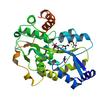
X-ray diffraction data for the Crystal structure of the putative periplasmic solute-binding protein from Campylobacter jejuni
X-ray diffraction data for the 1.55 Angstrom Crystal Structure of N-acetylmuramic acid 6-phosphate Etherase from Yersinia enterocolitica.
X-ray diffraction data for the Cycloalternan-degrading enzyme from Trueperella pyogenes in complex with cycloalternan
