6943 results
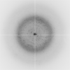
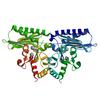
X-ray diffraction data for the Crystal structure of extragenic suppressor protein suhB from Bartonella henselae, native
First author:
T.E. Edwards
Resolution: 1.85 Å
R/Rfree: 0.19/0.22
Resolution: 1.85 Å
R/Rfree: 0.19/0.22
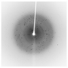
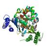
X-ray diffraction data for the Crystal structure of extragenic suppressor protein suhB from Bartonella henselae, via combined iodide SAD molecular replacement
First author:
J. Abendroth
Resolution: 2.05 Å
R/Rfree: 0.22/0.26
Resolution: 2.05 Å
R/Rfree: 0.22/0.26
X-ray diffraction data for the Crystal structure of beta-ketoacyl synthase from brucella melitensis

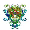
First author:
J. Abendroth
Resolution: 1.60 Å
R/Rfree: 0.13/0.16
Resolution: 1.60 Å
R/Rfree: 0.13/0.16
X-ray diffraction data for the Periplasmic domain of the risS sensor protein from Burkholderia pseudomallei, iodide phased at neutral pH
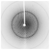
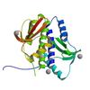
First author:
T.E. Edwards
Resolution: 2.30 Å
R/Rfree: 0.20/0.25
Resolution: 2.30 Å
R/Rfree: 0.20/0.25
X-ray diffraction data for the Periplasmic domain of the risS sensor protein from Burkholderia pseudomallei, barium phased at low pH
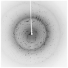
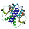
First author:
T.E. Edwards
Resolution: 1.90 Å
R/Rfree: 0.19/0.23
Resolution: 1.90 Å
R/Rfree: 0.19/0.23
X-ray diffraction data for the Periplasmic domain of the risS sensor protein from Burkholderia pseudomallei, iodide phased at low pH
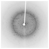
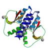
First author:
T.E. Edwards
Resolution: 1.90 Å
R/Rfree: 0.21/0.25
Resolution: 1.90 Å
R/Rfree: 0.21/0.25
X-ray diffraction data for the Crystal structure of deoxyuridine 5-triphosphate nucleotidohydrolase from Entamoeba histolytica
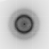
First author:
J. Abendroth
Resolution: 1.30 Å
R/Rfree: 0.13/0.16
Resolution: 1.30 Å
R/Rfree: 0.13/0.16
X-ray diffraction data for the Crystal structure of phosphoribosylamine-glycine ligase from Ehrlichia chaffeensis
First author:
Abendroth Seattle Structural Genomics Center for Infectious Disease (SSGCID)
Resolution: 2.15 Å
R/Rfree: 0.17/0.22
Resolution: 2.15 Å
R/Rfree: 0.17/0.22
X-ray diffraction data for the Crystal structure of inorganic pyrophosphatase from Ehrlichia chaffeensis
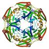
First author:
J. Abendroth
Resolution: 1.95 Å
R/Rfree: 0.21/0.26
Resolution: 1.95 Å
R/Rfree: 0.21/0.26
X-ray diffraction data for the Crystal structure of phosphoglyceromutase from Burkholderia Pseudomallei 1710B with bound malonic acid
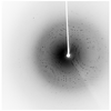
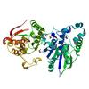
First author:
D.R. Davies
Resolution: 2.10 Å
R/Rfree: 0.17/0.20
Resolution: 2.10 Å
R/Rfree: 0.17/0.20