6907 results
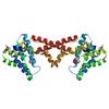
X-ray diffraction data for the Fragment of 7SK snRNA methylphosphate capping enzyme
First author:
H. Wu
Resolution: 2.55 Å
R/Rfree: 0.20/0.22
Resolution: 2.55 Å
R/Rfree: 0.20/0.22
First author:
K. Liu
Resolution: 2.30 Å
R/Rfree: 0.23/0.29
Resolution: 2.30 Å
R/Rfree: 0.23/0.29
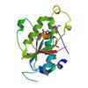
X-ray diffraction data for the Crystal structure of predicted HD superfamily hydrolase (104161995) from uncultured Thermotogales bacterium at 1.45 A resolution
First author:
Joint Center for Structural Genomics (JCSG)
Resolution: 1.45 Å
R/Rfree: 0.18/0.19
Resolution: 1.45 Å
R/Rfree: 0.18/0.19
First author:
K. Liu
Resolution: 2.01 Å
R/Rfree: 0.21/0.25
Resolution: 2.01 Å
R/Rfree: 0.21/0.25
First author:
K. Liu
Resolution: 1.90 Å
R/Rfree: 0.22/0.25
Resolution: 1.90 Å
R/Rfree: 0.22/0.25
First author:
K. Liu
Resolution: 1.95 Å
R/Rfree: 0.22/0.26
Resolution: 1.95 Å
R/Rfree: 0.22/0.26
First author:
M. Lei
Resolution: 2.30 Å
R/Rfree: 0.23/0.27
Resolution: 2.30 Å
R/Rfree: 0.23/0.27
First author:
M. Lei
Resolution: 2.30 Å
R/Rfree: 0.23/0.27
Resolution: 2.30 Å
R/Rfree: 0.23/0.27
First author:
M. Lei
Resolution: 1.84 Å
R/Rfree: 0.21/0.25
Resolution: 1.84 Å
R/Rfree: 0.21/0.25
First author:
K. Liu
Resolution: 2.05 Å
R/Rfree: 0.23/0.27
Resolution: 2.05 Å
R/Rfree: 0.23/0.27
First author:
K. Liu
Resolution: 2.65 Å
R/Rfree: 0.21/0.24
Resolution: 2.65 Å
R/Rfree: 0.21/0.24
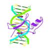
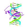
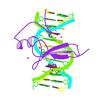
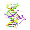
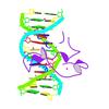
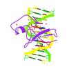
X-ray diffraction data for the MBD2 in complex with methylated DNA
First author:
K. Liu
Resolution: 2.15 Å
R/Rfree: 0.21/0.23
Resolution: 2.15 Å
R/Rfree: 0.21/0.23
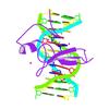
X-ray diffraction data for the Crystal structure of MBD2 complex with methylated CpG island
First author:
C. Bian
Resolution: 2.10 Å
R/Rfree: 0.20/0.22
Resolution: 2.10 Å
R/Rfree: 0.20/0.22
First author:
C. Xu
Resolution: 1.80 Å
R/Rfree: 0.23/0.25
Resolution: 1.80 Å
R/Rfree: 0.23/0.25
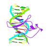
X-ray diffraction data for the Complex of MBD1-MBD and methylated DNA
First author:
K. Liu
Resolution: 2.25 Å
R/Rfree: 0.24/0.27
Resolution: 2.25 Å
R/Rfree: 0.24/0.27