393 results
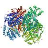
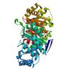
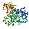
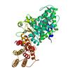
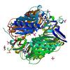
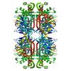
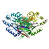
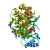
X-ray diffraction data for the 2.60 Angstrom resolution crystal structure of betaine aldehyde dehydrogenase (betB) H448F/Y450L double mutant from Staphylococcus aureus in complex with NAD+ and BME-free Cys289

First author:
A.S. Halavaty
Gene name: betB
Resolution: 2.60 Å
R/Rfree: 0.16/0.23
Gene name: betB
Resolution: 2.60 Å
R/Rfree: 0.16/0.23
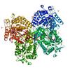
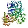
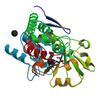
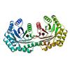
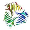
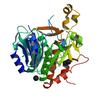
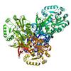
X-ray diffraction data for the 2.85 Angstrom resolution crystal structure of betaine aldehyde dehydrogenase (betB) H448F/P449M double mutant from Staphylococcus aureus in complex with NAD+ and BME-free Cys289
First author:
C. Chen
Gene name: betB
Resolution: 2.85 Å
R/Rfree: 0.16/0.21
Gene name: betB
Resolution: 2.85 Å
R/Rfree: 0.16/0.21
X-ray diffraction data for the Crystal Structure of K170M Mutant of Type I 3-Dehydroquinate Dehydratase (aroD) from Salmonella typhimurium LT2 in Non-Covalent Complex with Dehydroquinate.
First author:
S.H. Light
Gene name: aroD
Resolution: 1.60 Å
R/Rfree: 0.16/0.19
Gene name: aroD
Resolution: 1.60 Å
R/Rfree: 0.16/0.19
X-ray diffraction data for the Crystal structure of anabolic ornithine carbamoyltransferase from Vibrio vulnificus in complex with citrulline and inorganic phosphate
First author:
I.G. Shabalin
Resolution: 2.08 Å
R/Rfree: 0.16/0.19
Resolution: 2.08 Å
R/Rfree: 0.16/0.19
X-ray diffraction data for the Crystal structure of anabolic ornithine carbamoyltransferase from Vibrio vulnificus in complex with carbamoylphosphate and arginine
First author:
I.G. Shabalin
Resolution: 1.85 Å
R/Rfree: 0.15/0.19
Resolution: 1.85 Å
R/Rfree: 0.15/0.19
X-ray diffraction data for the Crystal structure of iron uptake ABC transporter substrate-binding protein PiaA from Streptococcus pneumoniae Canada MDR_19A bound to Bis-tris propane
First author:
P.J. Stogios
Resolution: 1.66 Å
R/Rfree: 0.17/0.19
Resolution: 1.66 Å
R/Rfree: 0.17/0.19
X-ray diffraction data for the Crystal structure of a putative Cas1 enzyme from Vibrio phage ICP1
First author:
A. Savchenko
Gene name: cas1
Resolution: 2.13 Å
R/Rfree: 0.17/0.21
Gene name: cas1
Resolution: 2.13 Å
R/Rfree: 0.17/0.21
X-ray diffraction data for the Crystal Structure of Type I 3-Dehydroquinate Dehydratase (aroD) from Salmonella typhimurium with close loop conformation.
First author:
S.H. Light
Gene name: aroD
Resolution: 1.90 Å
R/Rfree: 0.15/0.20
Gene name: aroD
Resolution: 1.90 Å
R/Rfree: 0.15/0.20
X-ray diffraction data for the 2.09 Angstrom resolution structure of a hypoxanthine-guanine phosphoribosyltransferase (hpt-1) from Bacillus anthracis str. 'Ames Ancestor' in complex with GMP
First author:
A.S. Halavaty
Gene name: hpt-1
Resolution: 2.09 Å
R/Rfree: 0.20/0.24
Gene name: hpt-1
Resolution: 2.09 Å
R/Rfree: 0.20/0.24
X-ray diffraction data for the 1.70 Angstrom resolution crystal structure of outer-membrane lipoprotein carrier protein (lolA) from Yersinia pestis CO92
First author:
A.S. Halavaty
Gene name: lolA
Resolution: 1.70 Å
R/Rfree: 0.20/0.24
Gene name: lolA
Resolution: 1.70 Å
R/Rfree: 0.20/0.24
X-ray diffraction data for the Crystal structure of spermidine N-acetyltransferase from Vibrio cholerae in complex with spermine
First author:
E.V. Filippova
Resolution: 1.85 Å
R/Rfree: 0.15/0.18
Resolution: 1.85 Å
R/Rfree: 0.15/0.18
X-ray diffraction data for the 1.95 Angstrom Resolution Crystal Structure of Epidermin Leader Peptide Processing Serine Protease (EpiP) S393A Mutant from Staphylococcus aureus
First author:
G. Minasov
Gene name: epiP
Resolution: 1.95 Å
R/Rfree: 0.17/0.20
Gene name: epiP
Resolution: 1.95 Å
R/Rfree: 0.17/0.20
X-ray diffraction data for the 2.4 Angstrom Crystal Structure of Dihydroorotase (pyrC) from Campylobacter jejuni.
First author:
G. Minasov
Gene name: pyrC
Resolution: 2.40 Å
R/Rfree: 0.19/0.25
Gene name: pyrC
Resolution: 2.40 Å
R/Rfree: 0.19/0.25
X-ray diffraction data for the 1.8 Angstrom resolution crystal structure of dihydroorotase (pyrC) from Salmonella enterica subsp. enterica serovar Typhimurium str. LT2
First author:
G. Minasov
Gene name: pyrC
Resolution: 1.80 Å
R/Rfree: 0.16/0.20
Gene name: pyrC
Resolution: 1.80 Å
R/Rfree: 0.16/0.20
X-ray diffraction data for the Crystal Structure of the 3-Dehydroquinate Dehydratase (aroD) from Salmonella typhimurium LT2 with Citrate Bound to the Active Site.
First author:
G. Minasov
Gene name: aroD
Resolution: 1.65 Å
R/Rfree: 0.16/0.20
Gene name: aroD
Resolution: 1.65 Å
R/Rfree: 0.16/0.20