420 results
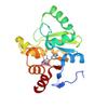
X-ray diffraction data for the PanDDA analysis group deposition -- Crystal structure of SARS-CoV-2 NSP3 macrodomain in complex with REAL250004627335
First author:
G.J. Correy
Resolution: 1.05 Å
R/Rfree: 0.14/0.16
Resolution: 1.05 Å
R/Rfree: 0.14/0.16
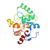
X-ray diffraction data for the PanDDA analysis group deposition -- Crystal structure of SARS-CoV-2 NSP3 macrodomain in complex with ZINC000833624464 - (R,R) and (S,S) isomers
First author:
G.J. Correy
Resolution: 1.05 Å
R/Rfree: 0.14/0.16
Resolution: 1.05 Å
R/Rfree: 0.14/0.16
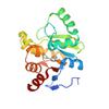
X-ray diffraction data for the PanDDA analysis group deposition -- Crystal structure of SARS-CoV-2 NSP3 macrodomain in complex with Z2479782408 - (R,S) isomer
First author:
G.J. Correy
Resolution: 1.05 Å
R/Rfree: 0.13/0.15
Resolution: 1.05 Å
R/Rfree: 0.13/0.15
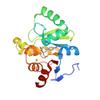
X-ray diffraction data for the PanDDA analysis group deposition -- Crystal structure of SARS-CoV-2 NSP3 macrodomain in complex with Z5021668601
First author:
G.J. Correy
Resolution: 1.05 Å
R/Rfree: 0.13/0.15
Resolution: 1.05 Å
R/Rfree: 0.13/0.15
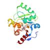
X-ray diffraction data for the PanDDA analysis group deposition -- Crystal structure of SARS-CoV-2 NSP3 macrodomain in complex with Z5016127255 - (R,R) and (S,S) isomers
First author:
G.J. Correy
Resolution: 1.05 Å
R/Rfree: 0.14/0.16
Resolution: 1.05 Å
R/Rfree: 0.14/0.16
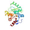
X-ray diffraction data for the PanDDA analysis group deposition -- Crystal structure of SARS-CoV-2 NSP3 macrodomain in complex with Z5010894431- (S,S) isomer
First author:
G.J. Correy
Resolution: 1.05 Å
R/Rfree: 0.13/0.15
Resolution: 1.05 Å
R/Rfree: 0.13/0.15
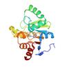
X-ray diffraction data for the PanDDA analysis group deposition -- Crystal structure of SARS-CoV-2 NSP3 macrodomain in complex with Z5030903496 - (R) isomer
First author:
G.J. Correy
Resolution: 1.05 Å
R/Rfree: 0.14/0.16
Resolution: 1.05 Å
R/Rfree: 0.14/0.16
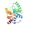
X-ray diffraction data for the PanDDA analysis group deposition -- Crystal structure of SARS-CoV-2 NSP3 macrodomain in complex with Z4914649780 - (S) isomer
First author:
G.J. Correy
Resolution: 1.05 Å
R/Rfree: 0.14/0.16
Resolution: 1.05 Å
R/Rfree: 0.14/0.16
X-ray diffraction data for the PanDDA analysis group deposition -- Crystal structure of SARS-CoV-2 NSP3 macrodomain in complex with Z5028367849 - (S) isomer
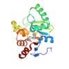
First author:
G.J. Correy
Resolution: 1.05 Å
R/Rfree: 0.14/0.16
Resolution: 1.05 Å
R/Rfree: 0.14/0.16
X-ray diffraction data for the PanDDA analysis group deposition -- Crystal structure of SARS-CoV-2 NSP3 macrodomain in complex with Z2689779890
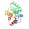
First author:
G.J. Correy
Resolution: 1.05 Å
R/Rfree: 0.13/0.15
Resolution: 1.05 Å
R/Rfree: 0.13/0.15
X-ray diffraction data for the PanDDA analysis group deposition -- Crystal structure of SARS-CoV-2 NSP3 macrodomain in complex with Z5010894388 - (R,R) and (S,S) isomers

First author:
G.J. Correy
Resolution: 1.05 Å
R/Rfree: 0.13/0.15
Resolution: 1.05 Å
R/Rfree: 0.13/0.15
X-ray diffraction data for the PanDDA analysis group deposition -- Crystal structure of SARS-CoV-2 NSP3 macrodomain in complex with Z5010894390 - (R,R) and (S,S) isomers
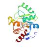
First author:
G.J. Correy
Resolution: 1.05 Å
R/Rfree: 0.12/0.14
Resolution: 1.05 Å
R/Rfree: 0.12/0.14
X-ray diffraction data for the PanDDA analysis group deposition -- Crystal structure of SARS-CoV-2 NSP3 macrodomain in complex with Z5010894395 - (R,R) and (S,S) isomers


First author:
G.J. Correy
Resolution: 1.05 Å
R/Rfree: 0.14/0.16
Resolution: 1.05 Å
R/Rfree: 0.14/0.16
X-ray diffraction data for the PanDDA analysis group deposition -- Crystal structure of SARS-CoV-2 NSP3 macrodomain in complex with Z5010894430 - (R,R) and (S,S) isomers
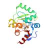
First author:
G.J. Correy
Resolution: 1.05 Å
R/Rfree: 0.14/0.16
Resolution: 1.05 Å
R/Rfree: 0.14/0.16
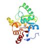
X-ray diffraction data for the PanDDA analysis group deposition -- Crystal structure of SARS-CoV-2 NSP3 macrodomain in complex with REAL250000548538 - (R) isomer
First author:
G.J. Correy
Resolution: 1.05 Å
R/Rfree: 0.14/0.15
Resolution: 1.05 Å
R/Rfree: 0.14/0.15
X-ray diffraction data for the PanDDA analysis group deposition -- Crystal structure of SARS-CoV-2 NSP3 macrodomain in complex with Z5010894392- (S,S) isomer
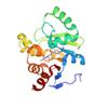
First author:
G.J. Correy
Resolution: 1.05 Å
R/Rfree: 0.13/0.15
Resolution: 1.05 Å
R/Rfree: 0.13/0.15
X-ray diffraction data for the PanDDA analysis group deposition -- Crystal structure of SARS-CoV-2 NSP3 macrodomain in complex with Z5010894382 - (R,R) and (S,S) isomers

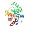
First author:
G.J. Correy
Resolution: 1.05 Å
R/Rfree: 0.13/0.15
Resolution: 1.05 Å
R/Rfree: 0.13/0.15
X-ray diffraction data for the PanDDA analysis group deposition -- Crystal structure of SARS-CoV-2 NSP3 macrodomain in complex with ZINC000450476923 - (S,R) isomer


First author:
G.J. Correy
Resolution: 1.01 Å
R/Rfree: 0.14/0.16
Resolution: 1.01 Å
R/Rfree: 0.14/0.16
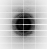
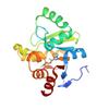
X-ray diffraction data for the PanDDA analysis group deposition -- Crystal structure of SARS-CoV-2 NSP3 macrodomain in complex with ZINCou000000a2Hm
X-ray diffraction data for the PanDDA analysis group deposition -- Crystal structure of SARS-CoV-2 NSP3 macrodomain in complex with ZINCn9000000uj1v
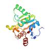
X-ray diffraction data for the PanDDA analysis group deposition -- Crystal structure of SARS-CoV-2 NSP3 macrodomain in complex with ZINCnu000001eLaQ
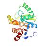
X-ray diffraction data for the PanDDA analysis group deposition -- Crystal structure of SARS-CoV-2 NSP3 macrodomain in complex with ZINCnt000006kx7L
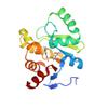
X-ray diffraction data for the PanDDA analysis group deposition -- Crystal structure of SARS-CoV-2 NSP3 macrodomain in complex with ZINCpE000000mAwk - (S) isomer
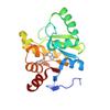
X-ray diffraction data for the PanDDA analysis group deposition -- Crystal structure of SARS-CoV-2 NSP3 macrodomain in complex with ZINCns000000RJoU
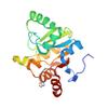
X-ray diffraction data for the PanDDA analysis group deposition -- Crystal structure of SARS-CoV-2 NSP3 macrodomain in complex with ZINCnz000004Qo8S
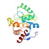
X-ray diffraction data for the PanDDA analysis group deposition -- Crystal structure of SARS-CoV-2 NSP3 macrodomain in complex with ZINClf00000cdzal
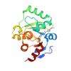
X-ray diffraction data for the PanDDA analysis group deposition -- Crystal structure of SARS-CoV-2 NSP3 macrodomain in complex with ZINCow000000AiWv - (R) isomer
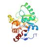
X-ray diffraction data for the PanDDA analysis group deposition -- Crystal structure of SARS-CoV-2 NSP3 macrodomain in complex with ZINCny000002NPIr
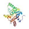
X-ray diffraction data for the PanDDA analysis group deposition -- Crystal structure of SARS-CoV-2 NSP3 macrodomain in complex with ZINC000681764827
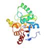
First author:
G.J. Correy
Resolution: 1.05 Å
R/Rfree: 0.13/0.15
Resolution: 1.05 Å
R/Rfree: 0.13/0.15
X-ray diffraction data for the PanDDA analysis group deposition -- Crystal structure of SARS-CoV-2 NSP3 macrodomain in complex with ZINClv000001jcNa - (r,r) isomer
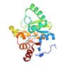
X-ray diffraction data for the PanDDA analysis group deposition -- Crystal structure of SARS-CoV-2 NSP3 macrodomain in complex with ZINCk500000doQ8X
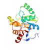
X-ray diffraction data for the PanDDA analysis group deposition -- Crystal structure of SARS-CoV-2 NSP3 macrodomain in complex with ZINCmk000007RhkC
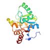
X-ray diffraction data for the PanDDA analysis group deposition -- Crystal structure of SARS-CoV-2 NSP3 macrodomain in complex with ZINCoD000001aHBe
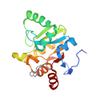
X-ray diffraction data for the PanDDA analysis group deposition -- Crystal structure of SARS-CoV-2 NSP3 macrodomain in complex with ZINCm4000007vvRA - (R,S) isomer
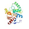
X-ray diffraction data for the PanDDA analysis group deposition -- Crystal structure of SARS-CoV-2 NSP3 macrodomain in complex with ZINCpv000006Li5M - (R,R) isomer
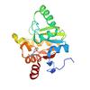
X-ray diffraction data for the PanDDA analysis group deposition -- Crystal structure of SARS-CoV-2 NSP3 macrodomain in complex with ZINCmr000000sTGN
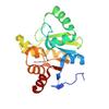
X-ray diffraction data for the PanDDA analysis group deposition -- Crystal structure of SARS-CoV-2 NSP3 macrodomain in complex with ZINCn500000bifGU
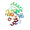
X-ray diffraction data for the PanDDA analysis group deposition -- Crystal structure of SARS-CoV-2 NSP3 macrodomain in complex with ZINCpx000006Mh4L - (S) isomer
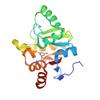
X-ray diffraction data for the PanDDA analysis group deposition -- Crystal structure of SARS-CoV-2 NSP3 macrodomain in complex with ZINCkk00000cjQyM - (R,S) isomer
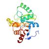
X-ray diffraction data for the PanDDA analysis group deposition -- Crystal structure of SARS-CoV-2 NSP3 macrodomain in complex with ZINCno00000broQT
X-ray diffraction data for the PanDDA analysis group deposition -- Crystal structure of SARS-CoV-2 NSP3 macrodomain in complex with Z5459166256 - (R,R) and (S,S) isomers
X-ray diffraction data for the PanDDA analysis group deposition -- Crystal structure of SARS-CoV-2 NSP3 macrodomain in complex with ZINCoj00000doMWF
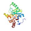
X-ray diffraction data for the PanDDA analysis group deposition -- Crystal structure of SARS-CoV-2 NSP3 macrodomain in complex with Z5198562519 - (R) and (S) isomers
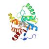
X-ray diffraction data for the PanDDA analysis group deposition -- Crystal structure of SARS-CoV-2 NSP3 macrodomain in complex with Z5010894382 - (R,S) isomer
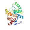
X-ray diffraction data for the PanDDA analysis group deposition -- Crystal structure of SARS-CoV-2 NSP3 macrodomain in complex with Z5459166291
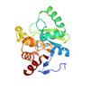
X-ray diffraction data for the PanDDA analysis group deposition -- Crystal structure of SARS-CoV-2 NSP3 macrodomain in complex with Z5459166285 - (R,R) and (S,S) isomers
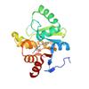
X-ray diffraction data for the PanDDA analysis group deposition -- Crystal structure of SARS-CoV-2 NSP3 macrodomain in complex with Z5459166300
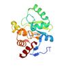
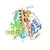
X-ray diffraction data for the Crystal structure of CYP124 in complex with inhibitor carbethoxyhexyl imidazole in the absence of glycerol (NoCryo)

First author:
S. Bukhdruker
Resolution: 1.15 Å
R/Rfree: 0.13/0.16
Resolution: 1.15 Å
R/Rfree: 0.13/0.16
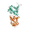
X-ray diffraction data for the Crystal structure of medical leech destabilase (high salt)
First author:
E. Marin
Resolution: 1.10 Å
R/Rfree: 0.12/0.16
Resolution: 1.10 Å
R/Rfree: 0.12/0.16
X-ray diffraction data for the Crystal Structure of Cystathionine beta lyase from Klebsiella aerogenes, PLP-Oxamate Adduct (C2 form)
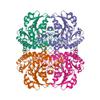
First author:
Seattle Structural Genomics Center for Infectious Disease (SSGCID)
Resolution: 1.10 Å
R/Rfree: 0.12/0.14
Resolution: 1.10 Å
R/Rfree: 0.12/0.14
X-ray diffraction data for the Crystal Structure of Dephospho-CoA kinase from Klebsiella aerogenes (P21 Form 1)
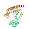
First author:
Seattle Structural Genomics Center for Infectious Disease (SSGCID)
Resolution: 1.15 Å
R/Rfree: 0.14/0.17
Resolution: 1.15 Å
R/Rfree: 0.14/0.17
X-ray diffraction data for the LOV2-DARPIN fusion : D4_deltaDARP
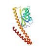
First author:
P. Mittl
Resolution: 1.10 Å
R/Rfree: 0.15/0.20
Resolution: 1.10 Å
R/Rfree: 0.15/0.20
X-ray diffraction data for the Reactive amide intermediate in sperm whale myoglobin mutant H64V V68A
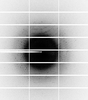

First author:
M. Tinzl
Resolution: 1.23 Å
R/Rfree: 0.15/0.18
Resolution: 1.23 Å
R/Rfree: 0.15/0.18
X-ray diffraction data for the Crystal Structure of UDP-N-acetylmuramoylalanine--D-glutamate ligase (MurD) from E. coli in complex with UMA and two inhibitor A19 molecules
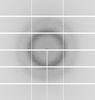
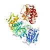
First author:
Seattle Structural Genomics Center for Infectious Disease (SSGCID)
Uniprot: P14900
Resolution: 1.23 Å
R/Rfree: 0.12/0.14
Uniprot: P14900
Resolution: 1.23 Å
R/Rfree: 0.12/0.14
X-ray diffraction data for the Crystal structure of peptidoglycan DL-endopeptidase CwlO
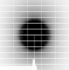
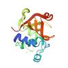
First author:
S. Tandukar
Resolution: 1.20 Å
R/Rfree: 0.15/0.16
Resolution: 1.20 Å
R/Rfree: 0.15/0.16
X-ray diffraction data for the High Resolution Crystal Structure of Putative Pterin Binding Protein (PruR) from Vibrio cholerae O1 biovar El Tor str. N16961 in Complex with Neopterin
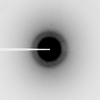
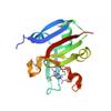
First author:
G. Minasov
Gene name: None
Resolution: 1.03 Å
R/Rfree: 0.13/0.15
Gene name: None
Resolution: 1.03 Å
R/Rfree: 0.13/0.15
X-ray diffraction data for the High Resolution Crystal Structure of Putative Pterin Binding Protein PruR (VV2_1280) from Vibrio vulnificus CMCP6
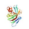
First author:
G. Minasov
Gene name: None
Resolution: 0.99 Å
R/Rfree: 0.12/0.14
Gene name: None
Resolution: 0.99 Å
R/Rfree: 0.12/0.14
X-ray diffraction data for the Crystal Structure of the dTDP-4-dehydrorhamnose Reductase from Streptococcus pneumoniae.
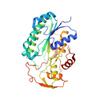
First author:
G. Minasov
Resolution: 1.00 Å
R/Rfree: 0.13/0.14
Resolution: 1.00 Å
R/Rfree: 0.13/0.14
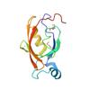
X-ray diffraction data for the Crystal Structure of the PDZ Domain of the Serine Peptidase HtrA from Streptococcus agalactiae.
First author:
G. Minasov
Gene name: htrA
Resolution: 0.97 Å
R/Rfree: 0.14/0.15
Gene name: htrA
Resolution: 0.97 Å
R/Rfree: 0.14/0.15
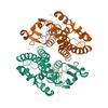
X-ray diffraction data for the Crystal structure of anthocyanin-related GSTF8 from Populus trichocarpa in complex with (-)-catechin and glutathione
X-ray diffraction data for the Crystal structure of ADP-ribose diphosphatase from Klebsiella pneumoniae (ADP bound, Orthorhombic P)
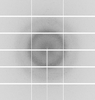
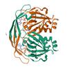
First author:
Seattle Structural Genomics Center for Infectious Disease (SSGCID) Seattle Structural Genomics Center for Infectious Disease
Uniprot: A0A0H3GVQ7
Resolution: 1.15 Å
R/Rfree: 0.14/0.16
Uniprot: A0A0H3GVQ7
Resolution: 1.15 Å
R/Rfree: 0.14/0.16
X-ray diffraction data for the Crystal structure of ADP-ribose diphosphatase from Klebsiella pneumoniae (5-O-phosphono-alpha-D-ribofuranose bound)
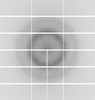
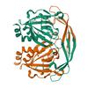
First author:
Seattle Structural Genomics Center for Infectious Disease (SSGCID) Seattle Structural Genomics Center for Infectious Disease
Uniprot: A0A0H3GVQ7
Resolution: 1.15 Å
R/Rfree: 0.13/0.15
Uniprot: A0A0H3GVQ7
Resolution: 1.15 Å
R/Rfree: 0.13/0.15
X-ray diffraction data for the X-ray Crystal Structure of Pseudoazurin Met16Gly variant
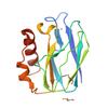
X-ray diffraction data for the The von Willebrand factor A domain of human capillary morphogenesis gene II, flexibly fused to the 1TEL crystallization chaperone, Thr-Val linker variant, at 1.2 Angstrom resolution

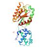
X-ray diffraction data for the Crystal structure of Apo Purine nucleoside phosphorylase from Trichomonas vaginalis (H32 form)

First author:
Seattle Structural Genomics Center for Infectious Disease (SSGCID) Seattle Structural Genomics Center for Infectious Disease
Uniprot: A2EU62
Resolution: 1.20 Å
R/Rfree: 0.11/0.13
Uniprot: A2EU62
Resolution: 1.20 Å
R/Rfree: 0.11/0.13
X-ray diffraction data for the Crystal structure of Purine nucleoside phosphorylase from Trichomonas vaginalis (mannose bound)

First author:
Seattle Structural Genomics Center for Infectious Disease (SSGCID) Seattle Structural Genomics Center for Infectious Disease
Uniprot: A2EU62
Resolution: 1.18 Å
R/Rfree: 0.13/0.15
Uniprot: A2EU62
Resolution: 1.18 Å
R/Rfree: 0.13/0.15
X-ray diffraction data for the Crystal structure of Purine nucleoside phosphorylase from Trichomonas vaginalis (adenosine and phosphate bound)
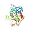
First author:
Seattle Structural Genomics Center for Infectious Disease (SSGCID) Seattle Structural Genomics Center for Infectious Disease
Uniprot: A2EU62
Resolution: 1.24 Å
R/Rfree: 0.13/0.15
Uniprot: A2EU62
Resolution: 1.24 Å
R/Rfree: 0.13/0.15
X-ray diffraction data for the Crystal structure of Purine nucleoside phosphorylase from Trichomonas vaginalis (inosine bound)

First author:
Seattle Structural Genomics Center for Infectious Disease (SSGCID) Seattle Structural Genomics Center for Infectious Disease
Resolution: 1.20 Å
R/Rfree: 0.13/0.16
Resolution: 1.20 Å
R/Rfree: 0.13/0.16
X-ray diffraction data for the A Stable Heterotrimeric Foldon with Minimum Mutation
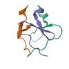
X-ray diffraction data for the Co-structure of the Fab of the anti-TIGIT Vibostolimab antibody with its antigen

X-ray diffraction data for the Crystal structure of Enterovirus A71 2A protease mutant C110A containing VP1-2A junction in the active site determined via sulfur phasing

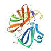
X-ray diffraction data for the CK1d in complex with IG130
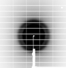
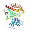
X-ray diffraction data for the Glucose/ xylose isomerase under 100 MPa
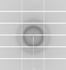
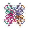
X-ray diffraction data for the Crystal Structure of a Phosphocarrier Protein HPr from Bacillus anthracis str. Ames
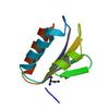
First author:
J.S. Brunzelle
Gene name: ptsH-2
Resolution: 1.15 Å
R/Rfree: 0.16/0.18
Gene name: ptsH-2
Resolution: 1.15 Å
R/Rfree: 0.16/0.18
X-ray diffraction data for the 0.95A Resolution Structure of a Histidine Triad Protein from Clostridium difficile
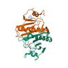
First author:
S.M. Anderson
Resolution: 0.95 Å
R/Rfree: 0.14/0.15
Resolution: 0.95 Å
R/Rfree: 0.14/0.15
X-ray diffraction data for the Structure of phosphotransferase enzyme II, A component from Yersinia pestis CO92 at 1.2 A resolution
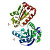
First author:
E.V. Filippova
Resolution: 1.20 Å
R/Rfree: 0.15/0.18
Resolution: 1.20 Å
R/Rfree: 0.15/0.18
X-ray diffraction data for the 1.05 Angstrom crystal structure of an amino acid ABC transporter substrate-binding protein AbpA from Streptococcus pneumoniae Canada MDR_19A bound to L-arginine
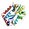
First author:
P.J. Stogios
Resolution: 1.05 Å
R/Rfree: 0.17/0.18
Resolution: 1.05 Å
R/Rfree: 0.17/0.18
X-ray diffraction data for the 1.2 Angstrom Crystal Structure of the Glutaredoxin 2 (grxB) from Salmonella typhimurium in complex with Glutathione
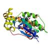
First author:
G. Minasov
Gene name: grxB
Resolution: 1.20 Å
R/Rfree: 0.12/0.14
Gene name: grxB
Resolution: 1.20 Å
R/Rfree: 0.12/0.14
X-ray diffraction data for the Crystal structure of PriA from Actinomyces urogenitalis
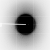
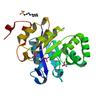
First author:
K. MICHALSKA
Resolution: 1.05 Å
R/Rfree: 0.12/0.14
Resolution: 1.05 Å
R/Rfree: 0.12/0.14
X-ray diffraction data for the 1.18 Angstrom resolution crystal structure of uncharacterized protein lmo1340 from Listeria monocytogenes EGD-e
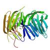
First author:
A.S. Halavaty
Gene name: -
Resolution: 1.18 Å
R/Rfree: 0.15/0.18
Gene name: -
Resolution: 1.18 Å
R/Rfree: 0.15/0.18
X-ray diffraction data for the 1.1 Angstrom Crystal Structure of Hypothetical Protein BA_2335 from Bacillus anthracis
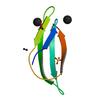
First author:
G. Minasov
Resolution: 1.10 Å
R/Rfree: 0.15/0.18
Resolution: 1.10 Å
R/Rfree: 0.15/0.18
X-ray diffraction data for the Crystal structure of VC2308 protein
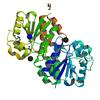
First author:
E. Niedzialkowska
Resolution: 1.16 Å
R/Rfree: 0.14/0.17
Resolution: 1.16 Å
R/Rfree: 0.14/0.17
X-ray diffraction data for the Crystal structure of a putative extracellular heme-binding protein (DESPIG_02683) from Desulfovibrio piger ATCC 29098 at 1.24 A resolution
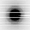
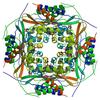
First author:
JOINT CENTER FOR STRUCTURAL GENOMICS (JCSG)
Resolution: 1.24 Å
R/Rfree: 0.13/0.15
Resolution: 1.24 Å
R/Rfree: 0.13/0.15
X-ray diffraction data for the Crystal structure of SusD superfamily protein (YP_001299712.1) from Bacteroides vulgatus ATCC 8482 at 1.13 A resolution
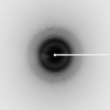
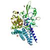
First author:
Joint Center for Structural Genomics (JCSG)
Resolution: 1.13 Å
R/Rfree: 0.12/0.14
Resolution: 1.13 Å
R/Rfree: 0.12/0.14
X-ray diffraction data for the PKD domain of M14-like peptidase from Thermoplasmatales archaeon SCGC AB-540-F20
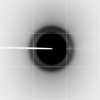
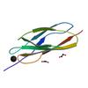
First author:
K. Michalska
Resolution: 1.23 Å
R/Rfree: 0.14/0.17
Resolution: 1.23 Å
R/Rfree: 0.14/0.17
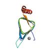
X-ray diffraction data for the Structure of the YrdA ferripyochelin binding protein from Salmonella enterica
First author:
S.M. Anderson
Gene name: yrdA
Resolution: 1.20 Å
R/Rfree: 0.14/0.16
Gene name: yrdA
Resolution: 1.20 Å
R/Rfree: 0.14/0.16
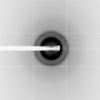
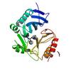
X-ray diffraction data for the New Delhi Metallo-beta-Lactamase-1 1.05 A structure Complexed with Hydrolyzed Ampicillin
First author:
Y. Kim
Resolution: 1.05 Å
R/Rfree: 0.13/0.16
Resolution: 1.05 Å
R/Rfree: 0.13/0.16
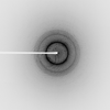
X-ray diffraction data for the Crystal structure of hypothetical protein with ketosteroid isomerase-like protein fold from Catenulispora acidiphila DSM 44928
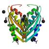
First author:
E.V. Filippova
Resolution: 1.15 Å
R/Rfree: 0.13/0.14
Resolution: 1.15 Å
R/Rfree: 0.13/0.14
X-ray diffraction data for the Crystal structure of TrpF from Jonesia denitrificans
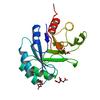
First author:
K. Michalska
Resolution: 1.09 Å
R/Rfree: 0.13/0.14
Resolution: 1.09 Å
R/Rfree: 0.13/0.14
X-ray diffraction data for the Fatty acid ABC transporter substrate-binding protein from Thermomonospora curvata
First author:
J. Osipiuk
Resolution: 1.15 Å
R/Rfree: 0.12/0.15
Resolution: 1.15 Å
R/Rfree: 0.12/0.15
X-ray diffraction data for the 1.03 Angstrom Crystal Structure of Q236A Mutant Type I Dehydroquinate Dehydratase (aroD) from Salmonella typhimurium
First author:
S.H. Light
Gene name: aroD
Resolution: 1.03 Å
R/Rfree: 0.14/0.16
Gene name: aroD
Resolution: 1.03 Å
R/Rfree: 0.14/0.16
X-ray diffraction data for the Crystal Structure of ThiJ/PfpI Domain Protein from Brachyspira murdochii
First author:
Y. Kim
Resolution: 1.12 Å
R/Rfree: 0.12/0.13
Resolution: 1.12 Å
R/Rfree: 0.12/0.13
X-ray diffraction data for the Crystal structure of a putative ornithine aminotransferase from Toxoplasma gondii ME49 in complex with pyrodoxal-5'-phosphate
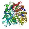
First author:
E.V. Filippova
Resolution: 1.20 Å
R/Rfree: 0.13/0.16
Resolution: 1.20 Å
R/Rfree: 0.13/0.16
X-ray diffraction data for the Crystal structure of transcriptional regulator VanUg, Form II
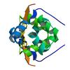
First author:
P.J. Stogios
Gene name: vanUG
Resolution: 1.12 Å
R/Rfree: 0.14/0.16
Gene name: vanUG
Resolution: 1.12 Å
R/Rfree: 0.14/0.16
X-ray diffraction data for the 1.0 Angstrom resolution crystal structure of the branched-chain amino acid transporter substrate binding protein LivJ from Streptococcus pneumoniae str. Canada MDR_19A in complex with Isoleucine
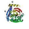
First author:
A.S. Halavaty
Resolution: 1.00 Å
R/Rfree: 0.13/0.15
Resolution: 1.00 Å
R/Rfree: 0.13/0.15
X-ray diffraction data for the Crystal structure of iron uptake ABC transporter substrate-binding protein PiuA from Streptococcus pneumoniae Canada MDR_19A
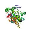
First author:
P.J. Stogios
Resolution: 1.13 Å
R/Rfree: 0.14/0.17
Resolution: 1.13 Å
R/Rfree: 0.14/0.17
X-ray diffraction data for the 1.02 Angstrom resolution crystal structure of 3-phosphoshikimate 1-carboxyvinyltransferase from Vibrio cholerae in complex with shikimate-3-phosphate (partially photolyzed) and glyphosate
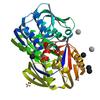
First author:
G. Minasov
Resolution: 1.02 Å
R/Rfree: 0.11/0.14
Resolution: 1.02 Å
R/Rfree: 0.11/0.14
X-ray diffraction data for the 1.1 Angstrom Crystal Structure of Putative Modulator of Drug Activity (MdaB) from Yersinia pestis CO92.
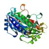
First author:
G. Minasov
Gene name: mdaB
Resolution: 1.10 Å
R/Rfree: 0.10/0.12
Gene name: mdaB
Resolution: 1.10 Å
R/Rfree: 0.10/0.12
X-ray diffraction data for the Structure of a NADH-dependent enoyl-ACP reductase from Acinetobacter baumannii in complex with NAD
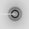
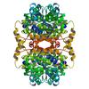
First author:
J. Abendroth
Resolution: 1.20 Å
R/Rfree: 0.13/0.14
Resolution: 1.20 Å
R/Rfree: 0.13/0.14
X-ray diffraction data for the X-ray crystal structure of thioredoxin from Mycobacterium avium
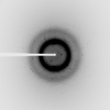
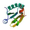
First author:
C.M. Lukacs
Resolution: 1.20 Å
R/Rfree: 0.16/0.18
Resolution: 1.20 Å
R/Rfree: 0.16/0.18