1764 results
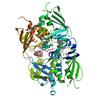
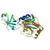
X-ray diffraction data for the Crystal structure of a conserved domain protein (SP_1775) from Streptococcus pneumoniae TIGR4 at 1.91 A resolution
First author:
JOINT CENTER FOR STRUCTURAL GENOMICS (JCSG)
Resolution: 1.91 Å
R/Rfree: 0.18/0.23
Resolution: 1.91 Å
R/Rfree: 0.18/0.23
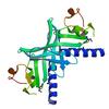
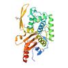
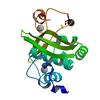
X-ray diffraction data for the Crystal structure of a xisi-like protein (npun_ar114) from nostoc punctiforme pcc 73102 at 2.30 A resolution
First author:
W.C. Hwang
Resolution: 2.30 Å
R/Rfree: 0.21/0.26
Resolution: 2.30 Å
R/Rfree: 0.21/0.26
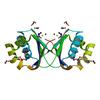
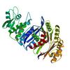
X-ray diffraction data for the Crystal structure of putative dioxygenase (YP_555069.1) from Burkholderia Xenovorans LB400 at 1.70 A resolution
First author:
Joint Center for Structural Genomics (JCSG)
Resolution: 1.70 Å
R/Rfree: 0.18/0.22
Resolution: 1.70 Å
R/Rfree: 0.18/0.22
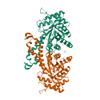
X-ray diffraction data for the Crystal structure of a putative nucleic acid binding protein (tm0693) from thermotoga maritima at 2.28 A resolution
First author:
Joint Center for Structural Genomics (JCSG)
Resolution: 2.28 Å
R/Rfree: 0.20/0.25
Resolution: 2.28 Å
R/Rfree: 0.20/0.25
X-ray diffraction data for the Crystal structure of a putative neuraminidase (BACOVA_03493) from Bacteroides ovatus ATCC 8483 at 1.74 A resolution
First author:
Joint Center for Structural Genomics (JCSG)
Resolution: 1.74 Å
R/Rfree: 0.17/0.21
Resolution: 1.74 Å
R/Rfree: 0.17/0.21
X-ray diffraction data for the Crystal structure of multidomain protein of unknown function with GGDEF-domain (NP_951600.1) from GEOBACTER SULFURREDUCENS at 1.95 A resolution
First author:
Joint Center for Structural Genomics (JCSG)
Resolution: 1.95 Å
R/Rfree: 0.18/0.23
Resolution: 1.95 Å
R/Rfree: 0.18/0.23
X-ray diffraction data for the Crystal structure of Endoglucanase (tm1049) from THERMOTOGA MARITIMA at 2.01 A resolution
First author:
Joint Center for Structural Genomics (JCSG)
Resolution: 2.01 Å
R/Rfree: 0.20/0.24
Resolution: 2.01 Å
R/Rfree: 0.20/0.24
X-ray diffraction data for the 1.75 Angstrom Resolution Crystal Structure of D-alanyl-D-alanine Endopeptidase from Enterobacter cloacae in Complex with Covalently Bound Boronic Acid
X-ray diffraction data for the 1.05 Angstrom Resolution Crystal Structure of UDP-N-acetylglucosamine 1-carboxyvinyltransferase from Acinetobacter baumannii in Covalently Bound Complex with (2R)-2-(phosphonooxy)propanoic Acid.
X-ray diffraction data for the 2.11 Angstrom resolution crystal structure of betaine aldehyde dehydrogenase (betB) P449M/Y450L double mutant from Staphylococcus aureus in complex with NAD+ and BME-modified Cys289


X-ray diffraction data for the 1.3 Angstrom Resolution Crystal Structure of UDP-N-acetylglucosamine 1-carboxyvinyltransferase from Streptococcus pneumoniae in Complex with (2R)-2-(phosphonooxy)propanoic acid.
X-ray diffraction data for the 2.85 Angstrom resolution crystal structure of betaine aldehyde dehydrogenase (betB) H448F/P449M double mutant from Staphylococcus aureus in complex with NAD+ and BME-free Cys289
First author:
A.S. Halavaty
Gene name: betB
Resolution: 2.85 Å
R/Rfree: 0.16/0.21
Gene name: betB
Resolution: 2.85 Å
R/Rfree: 0.16/0.21
X-ray diffraction data for the 1.70 Angstrom resolution crystal structure of outer-membrane lipoprotein carrier protein (lolA) from Yersinia pestis CO92
First author:
A.S. Halavaty
Gene name: lolA
Resolution: 1.70 Å
R/Rfree: 0.20/0.24
Gene name: lolA
Resolution: 1.70 Å
R/Rfree: 0.20/0.24
X-ray diffraction data for the 1.95 Angstrom Resolution Crystal Structure of Epidermin Leader Peptide Processing Serine Protease (EpiP) S393A Mutant from Staphylococcus aureus
First author:
G. Minasov
Gene name: epiP
Resolution: 1.95 Å
R/Rfree: 0.17/0.19
Gene name: epiP
Resolution: 1.95 Å
R/Rfree: 0.17/0.19
X-ray diffraction data for the 1.98 Angstrom resolution crystal structure of a hypoxanthine-guanine phosphoribosyltransferase (hpt-2) from Bacillus anthracis str. 'Ames Ancestor'

First author:
A.S. Halavaty
Gene name: hpt-2
Resolution: 1.98 Å
R/Rfree: 0.17/0.21
Gene name: hpt-2
Resolution: 1.98 Å
R/Rfree: 0.17/0.21
X-ray diffraction data for the 1.85 Angstrom resolution crystal structure of apo betaine aldehyde dehydrogenase (betB) G234S mutant from Staphylococcus aureus (IDP00699) with BME-free sulfinic acid form of Cys289
First author:
A.S. Halavaty
Gene name: betB
Resolution: 1.85 Å
R/Rfree: 0.15/0.18
Gene name: betB
Resolution: 1.85 Å
R/Rfree: 0.15/0.18
X-ray diffraction data for the Crystal structure of a streptavidin-like protein (BACEGG_01519) from Bacteroides eggerthii DSM 20697 at 1.25 A resolution

First author:
JOINT CENTER FOR STRUCTURAL GENOMICS (JCSG)
Resolution: 1.25 Å
R/Rfree: 0.13/0.15
Resolution: 1.25 Å
R/Rfree: 0.13/0.15
X-ray diffraction data for the Crystal structure of a dabb family protein with a ferredoxin-like fold (mll5499) from mesorhizobium loti maff303099 at 1.79 A resolution
First author:
Joint Center for Structural Genomics (JCSG)
Resolution: 1.79 Å
R/Rfree: 0.18/0.22
Resolution: 1.79 Å
R/Rfree: 0.18/0.22
X-ray diffraction data for the Crystal structure of a 5-keto-2-deoxygluconokinase (NCgl0155, Cgl0158) from Corynebacterium glutamicum ATCC 13032 KITASATO at 1.89 A resolution

First author:
Joint Center for Structural Genomics (JCSG)
Resolution: 1.89 Å
R/Rfree: 0.18/0.23
Resolution: 1.89 Å
R/Rfree: 0.18/0.23
X-ray diffraction data for the Crystal structure of a sec-c motif containing protein (psyc_2064) from psychrobacter arcticus at 1.75 A resolution
First author:
Joint Center for Structural Genomics (JCSG)
Resolution: 1.75 Å
R/Rfree: 0.19/0.24
Resolution: 1.75 Å
R/Rfree: 0.19/0.24
X-ray diffraction data for the 2.06 Angstrom resolution crystal structure of phosphomethylpyrimidine kinase (thiD)from Clostridium difficile 630
First author:
A.S. Halavaty
Gene name: thiD
Resolution: 2.06 Å
R/Rfree: 0.18/0.22
Gene name: thiD
Resolution: 2.06 Å
R/Rfree: 0.18/0.22
X-ray diffraction data for the 1.85 Angstrom Resolution Crystal Structure of Fructose-bisphosphate Aldolase (Fba) from Campylobacter jejuni
First author:
G. Minasov
Gene name: fba
Resolution: 1.85 Å
R/Rfree: 0.15/0.19
Gene name: fba
Resolution: 1.85 Å
R/Rfree: 0.15/0.19
X-ray diffraction data for the 2.05 Angstrom resolution crystal structure of a short chain dehydrogenase from Bacillus anthracis str. 'Ames Ancestor' in complex with NAD+
First author:
A.S. Halavaty
Resolution: 2.05 Å
R/Rfree: 0.16/0.19
Resolution: 2.05 Å
R/Rfree: 0.16/0.19
X-ray diffraction data for the 1.65 Angstrom resolution crystal structure of betaine aldehyde dehydrogenase (betB) from Staphylococcus aureus with BME-modified Cys289 and PEG molecule in active site
First author:
A.S. Halavaty
Gene name: betB
Resolution: 1.65 Å
R/Rfree: 0.14/0.16
Gene name: betB
Resolution: 1.65 Å
R/Rfree: 0.14/0.16
X-ray diffraction data for the 1.99 Angstrom resolution crystal structure of a short chain dehydrogenase from Bacillus anthracis str. 'Ames Ancestor'
First author:
A.S. Halavaty
Resolution: 1.99 Å
R/Rfree: 0.17/0.21
Resolution: 1.99 Å
R/Rfree: 0.17/0.21