6862 results
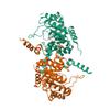
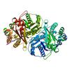
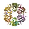
X-ray diffraction data for the Crystal structure of tertiary complex of glucosamine-6-phosphate deaminase from Vibrio cholerae with BETA-D-GLUCOSE-6-PHOSPHATE and FRUCTOSE-6-PHOSPHATE
First author:
C. Chang
Gene name: nagB
Resolution: 1.70 Å
R/Rfree: 0.14/0.17
Gene name: nagB
Resolution: 1.70 Å
R/Rfree: 0.14/0.17
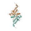
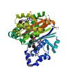
X-ray diffraction data for the Crystal structure of glucosamine-6-phosphate deaminase from Vibrio cholerae
First author:
N. Maltseva
Gene name: nagB
Resolution: 2.10 Å
R/Rfree: 0.20/0.25
Gene name: nagB
Resolution: 2.10 Å
R/Rfree: 0.20/0.25
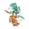
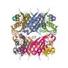
X-ray diffraction data for the Crystal Structure of the Inosine 5'-monophosphate Dehydrogenase, with a Short Internal Deletion of CBS Domain from Bacillus anthracis str. Ames
First author:
Y. Kim
Gene name: guaB
Resolution: 2.25 Å
R/Rfree: 0.21/0.23
Gene name: guaB
Resolution: 2.25 Å
R/Rfree: 0.21/0.23
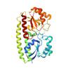
X-ray diffraction data for the Crystal Structure of the Inosine 5'-monophosphate Dehydrogenase, with a Internal Deletion of CBS Domain from Bacillus anthracis str. Ames complexed with P68
First author:
Y. Kim
Gene name: guaB
Resolution: 2.60 Å
R/Rfree: 0.19/0.25
Gene name: guaB
Resolution: 2.60 Å
R/Rfree: 0.19/0.25
X-ray diffraction data for the Crystal Structure of Carboxyvinyl-Carboxyphosphonate Phosphorylmutase from Bacillus anthracis
First author:
Y. Kim
Gene name: yqiQ
Resolution: 2.00 Å
R/Rfree: 0.14/0.19
Gene name: yqiQ
Resolution: 2.00 Å
R/Rfree: 0.14/0.19
X-ray diffraction data for the Crystal structure of putative periplasmic binding protein from Salmonella typhimurium LT2
First author:
C. Chang
Gene name: sitA
Resolution: 1.70 Å
R/Rfree: 0.17/0.21
Gene name: sitA
Resolution: 1.70 Å
R/Rfree: 0.17/0.21
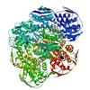
X-ray diffraction data for the Structure of UDP-N-acetylglucosamine 1-carboxyvinyltransferase from Vibrio cholerae in complex with substrate UDP-N-acetylglucosamine and the drug fosfomycin
First author:
B. Nocek
Resolution: 2.45 Å
R/Rfree: 0.18/0.23
Resolution: 2.45 Å
R/Rfree: 0.18/0.23
X-ray diffraction data for the Crystal Structure of Carboxyvinyl-Carboxyphosphonate Phosphorylmutase from Bacillus anthracis str. Ames Ancestor
First author:
Y. Kim
Gene name: yqiQ
Resolution: 2.50 Å
R/Rfree: 0.16/0.18
Gene name: yqiQ
Resolution: 2.50 Å
R/Rfree: 0.16/0.18
X-ray diffraction data for the Beta-ketoacyl-ACP synthase III -2 (FabH2) (C113A) from Vibrio Cholerae cocrystallized with octanoyl-CoA: hydrolzed ligand
First author:
J. Hou
Resolution: 2.00 Å
R/Rfree: 0.19/0.22
Resolution: 2.00 Å
R/Rfree: 0.19/0.22
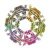
X-ray diffraction data for the Crystal structure of spermidine N-acetyltransferase from Vibrio cholerae in complex with CoA and spermine
First author:
E.V. Filippova
Resolution: 2.61 Å
R/Rfree: 0.18/0.24
Resolution: 2.61 Å
R/Rfree: 0.18/0.24
X-ray diffraction data for the Crystal structure putative autolysin from Listeria monocytogenes
First author:
C. Chang
Resolution: 2.15 Å
R/Rfree: 0.20/0.25
Resolution: 2.15 Å
R/Rfree: 0.20/0.25
X-ray diffraction data for the Crystal structure of spermidine N-acetyltransferase from Vibrio cholerae in complex with acetyl-CoA
First author:
E.V. Filippova
Resolution: 2.08 Å
R/Rfree: 0.17/0.24
Resolution: 2.08 Å
R/Rfree: 0.17/0.24
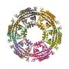
X-ray diffraction data for the Dodecameric structure of spermidine N-acetyltransferase from Vibrio cholerae in intermediate state
First author:
E.V. Filippova
Resolution: 2.50 Å
R/Rfree: 0.18/0.25
Resolution: 2.50 Å
R/Rfree: 0.18/0.25
X-ray diffraction data for the Crystal Structure of 4'-Phosphopantetheinyl Transferase AcpS from Vibrio cholerae O1 biovar eltor
First author:
Y. Kim
Gene name: acpS
Resolution: 1.85 Å
R/Rfree: 0.16/0.19
Gene name: acpS
Resolution: 1.85 Å
R/Rfree: 0.16/0.19
X-ray diffraction data for the Crystal structure of dihydroneopterin aldolase from Bacillus anthracis complexed with L-neopterin at 1.5 Angstroms resolution .
First author:
N. Maltseva
Gene name: folB
Resolution: 1.50 Å
R/Rfree: 0.14/0.16
Gene name: folB
Resolution: 1.50 Å
R/Rfree: 0.14/0.16
X-ray diffraction data for the Metal ABC transporter from Listeria monocytogenes with manganese
First author:
J. Osipiuk
Resolution: 1.79 Å
R/Rfree: 0.15/0.19
Resolution: 1.79 Å
R/Rfree: 0.15/0.19
X-ray diffraction data for the Metal ABC transporter from Listeria monocytogenes
First author:
J. Osipiuk
Resolution: 1.75 Å
R/Rfree: 0.17/0.20
Resolution: 1.75 Å
R/Rfree: 0.17/0.20
X-ray diffraction data for the 3.0 Angstrom Crystal Structure of 3-dehydroquinate Synthase (AroB) from Francisella tularensis in Complex with NAD.
First author:
G. Minasov
Gene name: aroB
Resolution: 3.00 Å
R/Rfree: 0.16/0.19
Gene name: aroB
Resolution: 3.00 Å
R/Rfree: 0.16/0.19
X-ray diffraction data for the Metal ABC transporter from Listeria monocytogenes with cadmium
First author:
J. Osipiuk
Resolution: 1.72 Å
R/Rfree: 0.17/0.21
Resolution: 1.72 Å
R/Rfree: 0.17/0.21
X-ray diffraction data for the Crystal structure of dihydroneopterin aldolase from Bacillus anthracis complex with 9-METHYLGUANINE
First author:
C. Chang
Gene name: folB
Resolution: 2.00 Å
R/Rfree: 0.19/0.23
Gene name: folB
Resolution: 2.00 Å
R/Rfree: 0.19/0.23
X-ray diffraction data for the Crystal Structure of Thiol-disulfide Oxidoreductase from Bacillus str. 'Ames Ancestor'
First author:
Y. Kim
Gene name: resA
Resolution: 1.35 Å
R/Rfree: 0.14/0.18
Gene name: resA
Resolution: 1.35 Å
R/Rfree: 0.14/0.18
X-ray diffraction data for the Crystal structure of putative aspartate racemase from Salmonella Typhimurium complexed with sulfate and potassium
First author:
N. Maltseva
Resolution: 1.70 Å
R/Rfree: 0.14/0.16
Resolution: 1.70 Å
R/Rfree: 0.14/0.16
X-ray diffraction data for the Crystal Structure of Putative Copper Homeostasis Protein CutC from Bacillus anthracis
First author:
Y. Kim
Resolution: 1.85 Å
R/Rfree: 0.18/0.21
Resolution: 1.85 Å
R/Rfree: 0.18/0.21
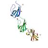
X-ray diffraction data for the Sortase B from Listeria monocytogenes.
First author:
J. Osipiuk
Resolution: 2.23 Å
R/Rfree: 0.21/0.25
Resolution: 2.23 Å
R/Rfree: 0.21/0.25
X-ray diffraction data for the Crystal Structure of Pantoate-beta-alanine Ligase from Francisella tularensis complex with SAM
First author:
C. Chang
Gene name: panC
Resolution: 2.40 Å
R/Rfree: 0.18/0.23
Gene name: panC
Resolution: 2.40 Å
R/Rfree: 0.18/0.23