6930 results
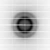
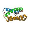
X-ray diffraction data for the Structure of the engineered metalloesterase MID1sc10 complexed with a phosphonate transition state analogue
First author:
P.R.E. Mittl
Resolution: 1.34 Å
R/Rfree: 0.13/0.17
Resolution: 1.34 Å
R/Rfree: 0.13/0.17
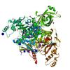
X-ray diffraction data for the Crystal Structure of Lysyl-tRNA Synthetase from Chlamydia trachomatis with complexed with L-lysine and Cladosporin

First author:
Seattle Structural Genomics Center for Infectious Disease (SSGCID) Seattle Structural Genomics Center for Infectious Disease
Resolution: 2.20 Å
R/Rfree: 0.16/0.21
Resolution: 2.20 Å
R/Rfree: 0.16/0.21
X-ray diffraction data for the Synthetic macromolecular crystallography diffraction image data generated to demonstrate the challenges of combining data from multiple crystals with indexing ambiguity in the context of heavy radiation damage - for more details see http://biorxiv.org/cgi/content/short/394965v1
First author:
James Holton
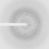
X-ray diffraction data for the Crystal structure of a full length elongation factor G (EF-G) from Rickettsia prowazekii
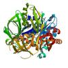
First author:
Seattle Structural Genomics Center for Infectious Disease (SSGCID)
Resolution: 2.40 Å
R/Rfree: 0.20/0.24
Resolution: 2.40 Å
R/Rfree: 0.20/0.24
X-ray diffraction data for the Crystal structure of prolyl-tRNA synthetase from Naegleria fowleri in complex with proline and adenosine monophophsphate (AMP)

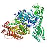
First author:
Seattle Structural Genomics Center for Infectious Disease (SSGCID)
Resolution: 2.00 Å
R/Rfree: 0.15/0.19
Resolution: 2.00 Å
R/Rfree: 0.15/0.19
X-ray diffraction data for the MBTD1 MBT repeats
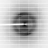
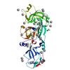
First author:
H. Zhang
Resolution: 1.95 Å
R/Rfree: 0.19/0.22
Resolution: 1.95 Å
R/Rfree: 0.19/0.22
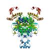
X-ray diffraction data for the Crystal structure of Histidine--tRNA ligase from Elizabethkingia sp. CCUG 26117
First author:
Seattle Structural Genomics Center for Infectious Disease (SSGCID)
Resolution: 2.10 Å
R/Rfree: 0.17/0.23
Resolution: 2.10 Å
R/Rfree: 0.17/0.23
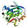
X-ray diffraction data for the MMP-13 in complex with the peptide IMISF
First author:
P. Mittl
Resolution: 1.71 Å
R/Rfree: 0.21/0.26
Resolution: 1.71 Å
R/Rfree: 0.21/0.26
X-ray diffraction data for the Crystal Structure of Glyceraldehyde-3-phosphate Dehydrogenase from Naegleria fowleri with bound NAD
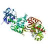
First author:
Seattle Structural Genomics Center for Infectious Disease (SSGCID) Seattle Structural Genomics Center for Infectious Disease
Resolution: 1.80 Å
R/Rfree: 0.17/0.20
Resolution: 1.80 Å
R/Rfree: 0.17/0.20
X-ray diffraction data for the CRYSTAL STRUCTURE OF ENOLASE FROM LEGIONELLA PNEUMOPHILA BOUND TO 2-PHOSPHOGLYCERIC ACID AND MAGNESIUM
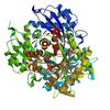
First author:
Seattle Structural Genomics Center for Infectious Disease (SSGCID)
Resolution: 1.85 Å
R/Rfree: 0.15/0.17
Resolution: 1.85 Å
R/Rfree: 0.15/0.17
X-ray diffraction data for the Crystal Structure of chromodomain of CBX8 in complex with inhibitor UNC3866
First author:
Y. Liu
Resolution: 1.18 Å
R/Rfree: 0.16/0.19
Resolution: 1.18 Å
R/Rfree: 0.16/0.19
X-ray diffraction data for the Crystal Structure of chromodomain of CBX7 in complex with inhibitor UNC3866
First author:
Y. Liu
Resolution: 1.60 Å
R/Rfree: 0.24/0.26
Resolution: 1.60 Å
R/Rfree: 0.24/0.26
X-ray diffraction data for the Crystal Structure of chromodomain of CBX4 in complex with inhibitor UNC3866
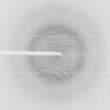
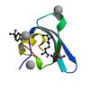
First author:
Y. Liu
Resolution: 1.81 Å
R/Rfree: 0.18/0.22
Resolution: 1.81 Å
R/Rfree: 0.18/0.22
X-ray diffraction data for the Crystal Structure of chromodomain of CBX2 in complex with inhibitor UNC3866
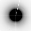
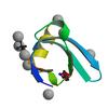
First author:
Y. Liu
Resolution: 1.80 Å
R/Rfree: 0.19/0.21
Resolution: 1.80 Å
R/Rfree: 0.19/0.21
X-ray diffraction data for the Crystal structure of a peptidase
First author:
A. Dong
Resolution: 2.50 Å
R/Rfree: 0.21/0.26
Resolution: 2.50 Å
R/Rfree: 0.21/0.26
X-ray diffraction data for the CRYSTAL STRUCTURE OF ENOLASE FROM LEGIONELLA PNEUMOPHILA BOUND TO PHOSPHATE AND MAGNESIUM
First author:
Seattle Structural Genomics Center for Infectious Disease (SSGCID)
Resolution: 1.90 Å
R/Rfree: 0.16/0.20
Resolution: 1.90 Å
R/Rfree: 0.16/0.20
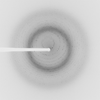
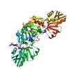
X-ray diffraction data for the Crystal Structure of Ribose-phosphate Pyrophosphokinase from Legionella pneumophila with bound AMP, ADP, and Ribose-5-Phosphate
First author:
Seattle Structural Genomics Center for Infectious Disease (SSGCID)
Resolution: 1.75 Å
R/Rfree: 0.15/0.18
Resolution: 1.75 Å
R/Rfree: 0.15/0.18
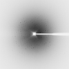
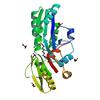
X-ray diffraction data for the The crystal structure of the Staphylococcus aureus Fatty acid Kinase (Fak) B1 protein loaded with 14-Methylhexadecanoic Acid (Anteiso C17:0) to 1.93 Angstrom resolution
First author:
M.G. Cuypers
Resolution: 1.93 Å
R/Rfree: 0.17/0.21
Resolution: 1.93 Å
R/Rfree: 0.17/0.21
X-ray diffraction data for the The crystal structure of the Staphylococcus aureus Fatty acid Kinase (Fak) B1 protein loaded with 12-Methyl Myristic Acid (C15:0) to 1.63 Angstrom resolution
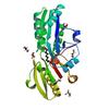
First author:
M.G. Cuypers
Resolution: 1.63 Å
R/Rfree: 0.16/0.20
Resolution: 1.63 Å
R/Rfree: 0.16/0.20
X-ray diffraction data for the The crystal structure of the Staphylococcus aureus Fatty acid Kinase (Fak) B1 protein loaded with Myristic acid (C14:0) to 1.78 Angstrom resolution
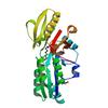
First author:
M.G. Cuypers
Resolution: 1.78 Å
R/Rfree: 0.15/0.20
Resolution: 1.78 Å
R/Rfree: 0.15/0.20
X-ray diffraction data for the The crystal structure of the Staphylococcus aureus Fatty acid Kinase (Fak) B1 protein loaded with palmitic acid to 1.83 Angstrom resolution
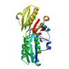
First author:
M.G. Cuypers
Resolution: 1.83 Å
R/Rfree: 0.16/0.19
Resolution: 1.83 Å
R/Rfree: 0.16/0.19
X-ray diffraction data for the The crystal structure of the bacteriophage T4 MotA C-terminal domain in complex with dsDNA reveals a novel protein-DNA recognition motif
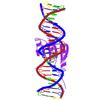
First author:
M.G. Cuypers
Resolution: 2.95 Å
R/Rfree: 0.22/0.25
Resolution: 2.95 Å
R/Rfree: 0.22/0.25
X-ray diffraction data for the Structure of USP5 zinc-finger ubiquitin binding domain co-crystallized with 3-(5-phenyl-1,3,4-oxadiazol-2-yl)propanoate
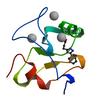
First author:
M.K. Mann
Resolution: 1.95 Å
R/Rfree: 0.19/0.24
Resolution: 1.95 Å
R/Rfree: 0.19/0.24
X-ray diffraction data for the Structure of USP5 zinc-finger ubiquitin binding domain co-crystallized with 4-(4-tert-butylphenyl)-4-oxobutanoate
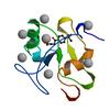
First author:
R.J. Harding
Resolution: 2.00 Å
R/Rfree: 0.21/0.25
Resolution: 2.00 Å
R/Rfree: 0.21/0.25
X-ray diffraction data for the Structure of the engineered metalloesterase MID1sc9
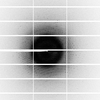
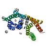
First author:
S. Studer
Resolution: 1.13 Å
R/Rfree: 0.15/0.19
Resolution: 1.13 Å
R/Rfree: 0.15/0.19