6890 results
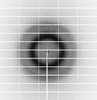
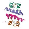
X-ray diffraction data for the Crystal structure of HLA-A0201 in complex with peptide LLWNGPMAVS

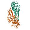
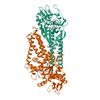
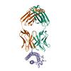
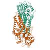
X-ray diffraction data for the Crystal structure of Cysteinyl-tRNA synthetase (CysRS) from Plasmodium falciparum (Hexagonal P form)
First author:
Seattle Structural Genomics Center for Infectious Disease (SSGCID)
Resolution: 2.87 Å
R/Rfree: 0.22/0.24
Resolution: 2.87 Å
R/Rfree: 0.22/0.24
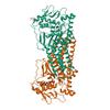
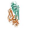
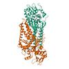
X-ray diffraction data for the Crystal structure of Cysteinyl-tRNA synthetase (CysRS) from Plasmodium falciparum in complex with ATP (long soak)

First author:
Seattle Structural Genomics Center for Infectious Disease (SSGCID)
Uniprot: Q8IJP3
Resolution: 2.80 Å
R/Rfree: 0.20/0.22
Uniprot: Q8IJP3
Resolution: 2.80 Å
R/Rfree: 0.20/0.22
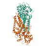
X-ray diffraction data for the Crystal structure of Cysteinyl-tRNA synthetase (CysRS) from Plasmodium falciparum in complex with AMP

First author:
Seattle Structural Genomics Center for Infectious Disease (SSGCID)
Uniprot: Q8IJP3
Resolution: 2.06 Å
R/Rfree: 0.18/0.20
Uniprot: Q8IJP3
Resolution: 2.06 Å
R/Rfree: 0.18/0.20
X-ray diffraction data for the Crystal structure of Cysteinyl-tRNA synthetase (CysRS) from Plasmodium falciparum in complex with GMP
First author:
Seattle Structural Genomics Center for Infectious Disease (SSGCID)
Uniprot: Q8IJP3
Resolution: 2.90 Å
R/Rfree: 0.19/0.22
Uniprot: Q8IJP3
Resolution: 2.90 Å
R/Rfree: 0.19/0.22
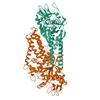
X-ray diffraction data for the Neutralizing monoclonal antibody Fab fragment bound to leptin
X-ray diffraction data for the Crystal structure of Cysteinyl-tRNA synthetase (CysRS) from Plasmodium falciparum in complex with O5'-(L-GLUTAMYL-SULFAMOYL)-ADENOSINE
First author:
Seattle Structural Genomics Center for Infectious Disease (SSGCID)
Uniprot: Q8IJP3
Resolution: 2.03 Å
R/Rfree: 0.18/0.20
Uniprot: Q8IJP3
Resolution: 2.03 Å
R/Rfree: 0.18/0.20
X-ray diffraction data for the Crystal structure of Cysteinyl-tRNA synthetase (CysRS) from Plasmodium falciparum in complex with Cysteine
First author:
Seattle Structural Genomics Center for Infectious Disease (SSGCID)
Uniprot: Q8IJP3
Resolution: 2.16 Å
R/Rfree: 0.19/0.22
Uniprot: Q8IJP3
Resolution: 2.16 Å
R/Rfree: 0.19/0.22
X-ray diffraction data for the Crystal structure of Cysteinyl-tRNA synthetase (CysRS) from Plasmodium falciparum in complex with ADP
First author:
Seattle Structural Genomics Center for Infectious Disease (SSGCID)
Uniprot: Q8IJP3
Resolution: 2.14 Å
R/Rfree: 0.18/0.20
Uniprot: Q8IJP3
Resolution: 2.14 Å
R/Rfree: 0.18/0.20
X-ray diffraction data for the Crystal structure of Cysteinyl-tRNA synthetase (CysRS) from Plasmodium falciparum in complex with AMP and Cysteine

First author:
Seattle Structural Genomics Center for Infectious Disease (SSGCID)
Uniprot: Q8IJP3
Resolution: 2.34 Å
R/Rfree: 0.19/0.22
Uniprot: Q8IJP3
Resolution: 2.34 Å
R/Rfree: 0.19/0.22
X-ray diffraction data for the Crystal structure of a Glyceraldehyde-3-phosphate dehydrogenase from Bordetella pertussis (monoclinic P form)
First author:
Seattle Structural Genomics Center for Infectious Disease (SSGCID) Seattle Structural Genomics Center for Infectious Disease
Uniprot: Q7VZB9
Resolution: 2.00 Å
R/Rfree: 0.17/0.20
Uniprot: Q7VZB9
Resolution: 2.00 Å
R/Rfree: 0.17/0.20
X-ray diffraction data for the Crystal Structure of Acetyl-CoA synthetase from Cryptococcus neoformans H99 in complex with inhibitor HGN-1310 (dd3-027)
First author:
Seattle Structural Genomics Center for Infectious Disease (SSGCID) Seattle Structural Genomics Center for Infectious Disease
Uniprot: J9VFT1
Resolution: 2.40 Å
R/Rfree: 0.18/0.22
Uniprot: J9VFT1
Resolution: 2.40 Å
R/Rfree: 0.18/0.22
X-ray diffraction data for the Rhombohedral crystalline form of human insulin complexed with m-cresol

X-ray diffraction data for the Crystal structure of Apo Cysteinyl-tRNA synthetase (CysRS) from Plasmodium falciparum (Orthrhombic P form)


First author:
Seattle Structural Genomics Center for Infectious Disease (SSGCID)
Uniprot: Q8IJP3
Resolution: 3.06 Å
R/Rfree: 0.23/0.29
Uniprot: Q8IJP3
Resolution: 3.06 Å
R/Rfree: 0.23/0.29
X-ray diffraction data for the Crystal structure of Cysteinyl-tRNA synthetase (CysRS) from Plasmodium falciparum in complex with 5'-Sulfamoyladenosine
First author:
Seattle Structural Genomics Center for Infectious Disease (SSGCID)
Uniprot: Q8IJP3
Resolution: 2.77 Å
R/Rfree: 0.19/0.23
Uniprot: Q8IJP3
Resolution: 2.77 Å
R/Rfree: 0.19/0.23