402 results
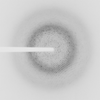
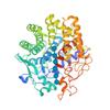
X-ray diffraction data for the Crystal structure of a CelR catalytic domain active site mutant with bound cellohexaose substrate
First author:
C.A. Bingman
Resolution: 1.90 Å
R/Rfree: 0.19/0.23
Resolution: 1.90 Å
R/Rfree: 0.19/0.23
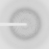
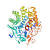
X-ray diffraction data for the Crystal structure of a CelR catalytic domain active site mutant with bound cellobiose product
First author:
C.A. Bingman
Resolution: 2.40 Å
R/Rfree: 0.19/0.22
Resolution: 2.40 Å
R/Rfree: 0.19/0.22
X-ray diffraction data for the Crystal Structure of K83A Mutant of Class D beta-lactamase from Clostridium difficile 630
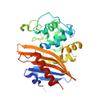
First author:
G. Minasov
Gene name:
Resolution: 1.88 Å
R/Rfree: 0.17/0.20
Gene name:
Resolution: 1.88 Å
R/Rfree: 0.17/0.20
X-ray diffraction data for the Crystals Structure of the Mutated Protease Domain of Botulinum Neurotoxin X (X4130B1).

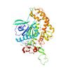
First author:
T.R. Blum
Resolution: 1.80 Å
R/Rfree: 0.15/0.18
Resolution: 1.80 Å
R/Rfree: 0.15/0.18
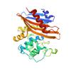
X-ray diffraction data for the Crystal Structure of C79A Mutant of Class D beta-lactamase from Clostridium difficile 630
First author:
G. Minasov
Gene name:
Resolution: 1.95 Å
R/Rfree: 0.18/0.21
Gene name:
Resolution: 1.95 Å
R/Rfree: 0.18/0.21
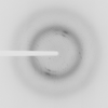
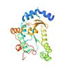
X-ray diffraction data for the 1.90 Angstrom Resolution Crystal Structure Phosphoadenosine Phosphosulfate Reductase (CysH) from Vibrio vulnificus
First author:
G. Minasov
Gene name: cysH
Resolution: 1.90 Å
R/Rfree: 0.17/0.21
Gene name: cysH
Resolution: 1.90 Å
R/Rfree: 0.17/0.21
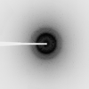
X-ray diffraction data for the High Resolution Crystal Structure of Putative Pterin Binding Protein PruR (VV2_1280) from Vibrio vulnificus CMCP6
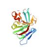
First author:
G. Minasov
Gene name: None
Resolution: 0.99 Å
R/Rfree: 0.12/0.14
Gene name: None
Resolution: 0.99 Å
R/Rfree: 0.12/0.14
X-ray diffraction data for the Crystal Structure of the DNA-binding Transcriptional Repressor DeoR from Escherichia coli str. K-12
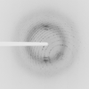
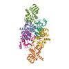
First author:
G. Minasov
Resolution: 1.75 Å
R/Rfree: 0.16/0.18
Resolution: 1.75 Å
R/Rfree: 0.16/0.18
X-ray diffraction data for the Crystal Structure of Putative Universal Stress Protein from Pseudomonas aeruginosa UCBPP-PA14
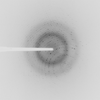
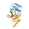
First author:
G. Minasov
Gene name: uspA
Resolution: 1.25 Å
R/Rfree: 0.13/0.16
Gene name: uspA
Resolution: 1.25 Å
R/Rfree: 0.13/0.16
X-ray diffraction data for the Crystal Structure of the C-terminal Fragment of AAA ATPase from Streptococcus pneumoniae.
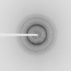
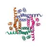
First author:
G. Minasov
Resolution: 1.30 Å
R/Rfree: 0.12/0.14
Resolution: 1.30 Å
R/Rfree: 0.12/0.14