1160 results
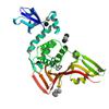
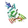
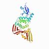
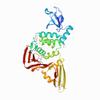
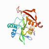
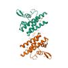
X-ray diffraction data for the The crystal structure of Papain-Like Protease of SARS CoV-2 in complex with PLP_Snyder441 inhibitor
First author:
J. Osipiuk
Resolution: 1.93 Å
R/Rfree: 0.19/0.21
Resolution: 1.93 Å
R/Rfree: 0.19/0.21
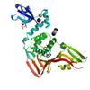
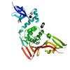
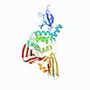
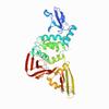
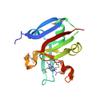
X-ray diffraction data for the The crystal structure of Papain-Like Protease of SARS CoV-2 , C111S mutant, in complex with PLP_Snyder530 inhibitor
First author:
J. Osipiuk
Resolution: 2.05 Å
R/Rfree: 0.19/0.20
Resolution: 2.05 Å
R/Rfree: 0.19/0.20
X-ray diffraction data for the The crystal structure of Papain-Like Protease of SARS CoV-2 , C111S mutant, in complex with PLP_Snyder457 inhibitor
First author:
J. Osipiuk
Resolution: 2.09 Å
R/Rfree: 0.19/0.20
Resolution: 2.09 Å
R/Rfree: 0.19/0.20
X-ray diffraction data for the The crystal structure of Papain-Like Protease of SARS CoV-2 , C111S mutant, in complex with PLP_Snyder495 inhibitor
First author:
J. Osipiuk
Resolution: 1.95 Å
R/Rfree: 0.17/0.19
Resolution: 1.95 Å
R/Rfree: 0.17/0.19
X-ray diffraction data for the The crystal structure of Papain-Like Protease of SARS CoV-2, C111S mutant, in complex with PLP_Snyder496 inhibitor

First author:
J. Osipiuk
Resolution: 2.00 Å
R/Rfree: 0.18/0.21
Resolution: 2.00 Å
R/Rfree: 0.18/0.21
X-ray diffraction data for the The crystal structure of Papain-Like Protease of SARS CoV-2, C111S mutant, in complex with PLP_Snyder494 inhibitor
First author:
J. Osipiuk
Resolution: 2.02 Å
R/Rfree: 0.18/0.20
Resolution: 2.02 Å
R/Rfree: 0.18/0.20
X-ray diffraction data for the The crystal structure of Papain-Like Protease of SARS CoV-2 in complex with PLP_Snyder496 inhibitor
First author:
J. Osipiuk
Resolution: 2.58 Å
R/Rfree: 0.19/0.21
Resolution: 2.58 Å
R/Rfree: 0.19/0.21
X-ray diffraction data for the The crystal structure of Papain-Like Protease of SARS CoV-2, C111S mutant, in complex with PLP_Snyder441 inhibitor
First author:
J. Osipiuk
Resolution: 2.72 Å
R/Rfree: 0.21/0.25
Resolution: 2.72 Å
R/Rfree: 0.21/0.25
X-ray diffraction data for the Structure of a GNAT superfamily PA3944 acetyltransferase in complex with zinc
First author:
M.P. Czub
Resolution: 2.00 Å
R/Rfree: 0.20/0.25
Resolution: 2.00 Å
R/Rfree: 0.20/0.25
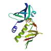
X-ray diffraction data for the High Resolution Crystal Structure of Putative Pterin Binding Protein (PruR) from Vibrio cholerae O1 biovar El Tor str. N16961 in Complex with Neopterin
First author:
G. Minasov
Gene name: None
Resolution: 1.03 Å
R/Rfree: 0.13/0.15
Gene name: None
Resolution: 1.03 Å
R/Rfree: 0.13/0.15
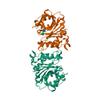
X-ray diffraction data for the High Resolution Crystal Structure of the DNA-binding Domain from the Sensor Histidine Kinase ChiS from Vibrio cholerae
First author:
G. Minasov
Resolution: 1.28 Å
R/Rfree: 0.14/0.18
Resolution: 1.28 Å
R/Rfree: 0.14/0.18
X-ray diffraction data for the 2.9 Angstrom Crystal Structure of Putative Exotoxin 3 from Staphylococcus aureus.
First author:
G. Minasov
Resolution: 2.90 Å
R/Rfree: 0.23/0.28
Resolution: 2.90 Å
R/Rfree: 0.23/0.28
X-ray diffraction data for the 2.76 Angstrom Crystal Structure of a Putative Glucose-1-Phosphate Thymidylyltransferase from Bacillus anthracis in Complex with a Sucrose.
First author:
G. Minasov
Resolution: 2.76 Å
R/Rfree: 0.17/0.21
Resolution: 2.76 Å
R/Rfree: 0.17/0.21
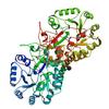
X-ray diffraction data for the 1.8 Angstrom Resolution Crystal Structure of Dihydroorotase (pyrc) from Salmonella enterica subsp. enterica serovar Typhimurium str. LT2.
First author:
A.Halavaty 'G.Minasov
Gene name: pyrC
Resolution: 1.80 Å
R/Rfree: 0.17/0.20
Gene name: pyrC
Resolution: 1.80 Å
R/Rfree: 0.17/0.20
X-ray diffraction data for the Crystal Structure of a Phosphocarrier Protein HPr from Bacillus anthracis str. Ames
First author:
J.S. Brunzelle
Gene name: ptsH-2
Resolution: 1.15 Å
R/Rfree: 0.16/0.18
Gene name: ptsH-2
Resolution: 1.15 Å
R/Rfree: 0.16/0.18