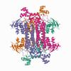
402 results
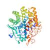
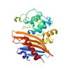
X-ray diffraction data for the Structure of RND efflux system, outer membrane lipoprotein, NodT family from Burkholderia mallei ATCC 23344
First author:
P.S. Horanyi
Resolution: 2.35 Å
Resolution: 2.35 Å
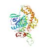
X-ray diffraction data for the Crystal Structure of Glucosamine-1-phosphate N-acetyltransferase from Stenotrophomonas maltophilia K279a
First author:
Seattle Structural Genomics Center for Infectious Disease (SSGCID)
Resolution: 2.90 Å
R/Rfree: 0.17/0.21
Resolution: 2.90 Å
R/Rfree: 0.17/0.21
X-ray diffraction data for the Crystal structure of a refolded head domain hemagglutinin HA from Influenza A virus A/Fort Monmouth/1/1947
First author:
Seattle Structural Genomics Center for Infectious Disease (SSGCID)
Resolution: 2.00 Å
R/Rfree: 0.19/0.23
Resolution: 2.00 Å
R/Rfree: 0.19/0.23
X-ray diffraction data for the Structure of E. coli Transcriptional Regulator RutR with bound uracil
First author:
D.R. Cooper
Resolution: 1.70 Å
R/Rfree: 0.12/0.17
Resolution: 1.70 Å
R/Rfree: 0.12/0.17
X-ray diffraction data for the Crystal Structure of Fumarate hydratase class II from Elizabethkingia anophelis NUHP1
First author:
Seattle Structural Genomics Center for Infectious Disease (SSGCID)
Resolution: 1.25 Å
R/Rfree: 0.12/0.15
Resolution: 1.25 Å
R/Rfree: 0.12/0.15
X-ray diffraction data for the Crystal Structure of aspartate-semialdehyde dehydrogenase from Acinetobacter baumannii
First author:
Seattle Structural Genomics Center for Infectious Disease (SSGCID)
Resolution: 1.80 Å
R/Rfree: 0.14/0.17
Resolution: 1.80 Å
R/Rfree: 0.14/0.17
X-ray diffraction data for the Crystal structure of a CelR catalytic domain active site mutant with bound cellohexaose substrate

First author:
C.A. Bingman
Resolution: 1.90 Å
R/Rfree: 0.19/0.23
Resolution: 1.90 Å
R/Rfree: 0.19/0.23
X-ray diffraction data for the Crystal structure of a CelR catalytic domain active site mutant with bound cellobiose product
First author:
C.A. Bingman
Resolution: 2.40 Å
R/Rfree: 0.19/0.22
Resolution: 2.40 Å
R/Rfree: 0.19/0.22
X-ray diffraction data for the Crystal Structure of K83A Mutant of Class D beta-lactamase from Clostridium difficile 630
First author:
G. Minasov
Gene name:
Resolution: 1.88 Å
R/Rfree: 0.17/0.20
Gene name:
Resolution: 1.88 Å
R/Rfree: 0.17/0.20
X-ray diffraction data for the Crystals Structure of the Mutated Protease Domain of Botulinum Neurotoxin X (X4130B1).

First author:
T.R. Blum
Resolution: 1.80 Å
R/Rfree: 0.15/0.18
Resolution: 1.80 Å
R/Rfree: 0.15/0.18
X-ray diffraction data for the Crystal Structure of C79A Mutant of Class D beta-lactamase from Clostridium difficile 630
First author:
G. Minasov
Gene name:
Resolution: 1.95 Å
R/Rfree: 0.18/0.21
Gene name:
Resolution: 1.95 Å
R/Rfree: 0.18/0.21
X-ray diffraction data for the 1.90 Angstrom Resolution Crystal Structure Phosphoadenosine Phosphosulfate Reductase (CysH) from Vibrio vulnificus
First author:
G. Minasov
Gene name: cysH
Resolution: 1.90 Å
R/Rfree: 0.17/0.21
Gene name: cysH
Resolution: 1.90 Å
R/Rfree: 0.17/0.21