1438 results
X-ray diffraction data for the Crystal Structure UTP--glucose-1-phosphate uridylyltransferase from Bordetella pertussis in complex with UTP
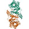
First author:
Seattle Structural Genomics Center for Infectious Disease (SSGCID)
Uniprot: Q7VTV0
Resolution: 1.82 Å
R/Rfree: 0.20/0.23
Uniprot: Q7VTV0
Resolution: 1.82 Å
R/Rfree: 0.20/0.23
X-ray diffraction data for the Crystal structure of Cysteinyl-tRNA synthetase (CysRS) from Plasmodium falciparum in complex with ADP
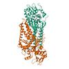
First author:
Seattle Structural Genomics Center for Infectious Disease (SSGCID)
Uniprot: Q8IJP3
Resolution: 2.14 Å
R/Rfree: 0.18/0.20
Uniprot: Q8IJP3
Resolution: 2.14 Å
R/Rfree: 0.18/0.20
X-ray diffraction data for the Crystal structure of Cysteinyl-tRNA synthetase (CysRS) from Plasmodium falciparum in complex with GMP
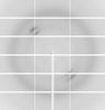
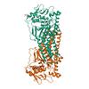
First author:
Seattle Structural Genomics Center for Infectious Disease (SSGCID)
Uniprot: Q8IJP3
Resolution: 2.90 Å
R/Rfree: 0.19/0.22
Uniprot: Q8IJP3
Resolution: 2.90 Å
R/Rfree: 0.19/0.22
X-ray diffraction data for the Crystal structure of Cysteinyl-tRNA synthetase (CysRS) from Plasmodium falciparum in complex with AMP

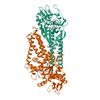
First author:
Seattle Structural Genomics Center for Infectious Disease (SSGCID)
Uniprot: Q8IJP3
Resolution: 2.06 Å
R/Rfree: 0.18/0.20
Uniprot: Q8IJP3
Resolution: 2.06 Å
R/Rfree: 0.18/0.20
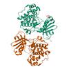
X-ray diffraction data for the Crystal Structure of serine/threonine-protein kinase (AEK1) from Trypanosoma brucei
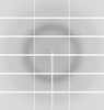
First author:
Seattle Structural Genomics Center for Infectious Disease (SSGCID)
Uniprot: Q582V7
Resolution: 2.05 Å
R/Rfree: 0.19/0.23
Uniprot: Q582V7
Resolution: 2.05 Å
R/Rfree: 0.19/0.23
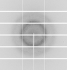
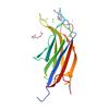
X-ray diffraction data for the Crystal structure of a calcium bound C2 domain containing protein from Trichomonas vaginalis (P61 form)
First author:
Seattle Structural Genomics Center for Infectious Disease (SSGCID)
Uniprot: A2EZR3
Resolution: 1.54 Å
R/Rfree: 0.17/0.20
Uniprot: A2EZR3
Resolution: 1.54 Å
R/Rfree: 0.17/0.20
X-ray diffraction data for the Crystal structure of a malonate bound C2 domain containing protein from Trichomonas vaginalis (P21 form)
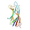
First author:
Seattle Structural Genomics Center for Infectious Disease (SSGCID)
Uniprot: A2EZR3
Resolution: 1.30 Å
R/Rfree: 0.14/0.17
Uniprot: A2EZR3
Resolution: 1.30 Å
R/Rfree: 0.14/0.17
X-ray diffraction data for the Crystal structure of a calcium bound C2 domain containing protein from Trichomonas vaginalis (P21 form)
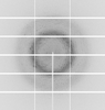
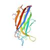
First author:
Seattle Structural Genomics Center for Infectious Disease (SSGCID)
Uniprot: A2EZR3
Resolution: 1.51 Å
R/Rfree: 0.19/0.21
Uniprot: A2EZR3
Resolution: 1.51 Å
R/Rfree: 0.19/0.21
X-ray diffraction data for the Crystal Structure of cyclophilin B, from Brugia malayi (K5H/S166A mutant)
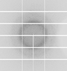
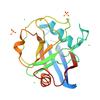
First author:
Seattle Structural Genomics Center for Infectious Disease (SSGCID)
Uniprot: A0A0J9XUF2
Resolution: 1.25 Å
R/Rfree: 0.11/0.13
Uniprot: A0A0J9XUF2
Resolution: 1.25 Å
R/Rfree: 0.11/0.13
X-ray diffraction data for the Crystal Structure of ribosomal large subunit pseudouridine synthase D from Neisseria gonorrhoea
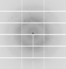
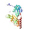
First author:
Seattle Structural Genomics Center for Infectious Disease (SSGCID)
Uniprot: B4RJW3
Resolution: 1.91 Å
R/Rfree: 0.17/0.19
Uniprot: B4RJW3
Resolution: 1.91 Å
R/Rfree: 0.17/0.19
X-ray diffraction data for the Crystal structure of a Phosphoribosylaminoimidazole carboxylase from Burkholderia xenovorans (Apo, Orthorhombic P form)
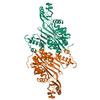
First author:
Seattle Structural Genomics Center for Infectious Disease (SSGCID)
Uniprot: Q13UJ9
Resolution: 2.05 Å
R/Rfree: 0.18/0.23
Uniprot: Q13UJ9
Resolution: 2.05 Å
R/Rfree: 0.18/0.23
X-ray diffraction data for the Crystal Structure of Histone-lysine N-methyltransferase from Leishmania major in complex with ATP
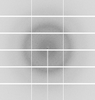
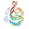
First author:
Seattle Structural Genomics Center for Infectious Disease (SSGCID)
Uniprot: Q4QIU2
Resolution: 1.96 Å
R/Rfree: 0.23/0.27
Uniprot: Q4QIU2
Resolution: 1.96 Å
R/Rfree: 0.23/0.27
X-ray diffraction data for the Crystal Structure of Histone-lysine N-methyltransferase from Leishmania major in complex with AMP
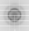
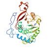
First author:
Seattle Structural Genomics Center for Infectious Disease (SSGCID)
Uniprot: Q4QIU2
Resolution: 1.80 Å
R/Rfree: 0.18/0.22
Uniprot: Q4QIU2
Resolution: 1.80 Å
R/Rfree: 0.18/0.22
X-ray diffraction data for the Crystal structure of an adenylate kinase from Leishmania major (Tetragonal form, ADP and AMP bound)
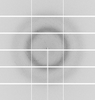
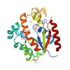
First author:
Seattle Structural Genomics Center for Infectious Disease (SSGCID)
Uniprot: Q4Q1U0
Resolution: 1.40 Å
R/Rfree: 0.15/0.19
Uniprot: Q4Q1U0
Resolution: 1.40 Å
R/Rfree: 0.15/0.19
X-ray diffraction data for the Crystal structure of phosphoprotein/Protein P/Protein M1 residues 69-297 from Rabies virus reveals degradation to C-terminal domain only
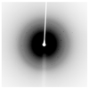
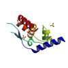
First author:
T.E. Edwards
Resolution: 2.20 Å
R/Rfree: 0.17/0.23
Resolution: 2.20 Å
R/Rfree: 0.17/0.23
X-ray diffraction data for the Crystal structure of nucleoside diphosphate kinase from Giardia lamblia featuring a disordered dinucleotide binding site
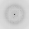
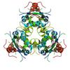
First author:
T.E. Edwards
Resolution: 2.65 Å
R/Rfree: 0.23/0.27
Resolution: 2.65 Å
R/Rfree: 0.23/0.27
X-ray diffraction data for the Crystal structure of Glutamate-1-semialdehyde 2,1- aminomutase (GSA) from Pseudomonas aeruginosa
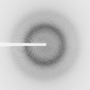
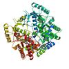
First author:
Delker Seattle Structural Genomics Center for Infectious Disease (SSGCID)
Resolution: 1.75 Å
R/Rfree: 0.15/0.18
Resolution: 1.75 Å
R/Rfree: 0.15/0.18
X-ray diffraction data for the Crystal Structure of Prolyl-tRNA Synthetase from Cryptosporidium parvum complexed with L-Proline and AMP
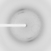
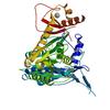
First author:
Dranow Seattle Structural Genomics Center for Infectious Disease (SSGCID)
Resolution: 2.80 Å
R/Rfree: 0.21/0.24
Resolution: 2.80 Å
R/Rfree: 0.21/0.24
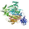
X-ray diffraction data for the Crystal Structure of Lysyl-tRNA Synthetase from Cryptosporidium parvum complexed with L-lysine and cladosporin
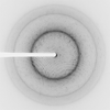
First author:
Dranow Seattle Structural Genomics Center for Infectious Disease (SSGCID)
Resolution: 1.90 Å
R/Rfree: 0.20/0.25
Resolution: 1.90 Å
R/Rfree: 0.20/0.25
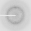
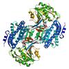
X-ray diffraction data for the Crystal structure of a Putative aldehyde dehydrogenase family protein Burkholderia cenocepacia J2315 in complex with partially reduced NADH
First author:
Seattle Structural Genomics Center for Infectious Disease (SSGCID)
Resolution: 2.10 Å
R/Rfree: 0.15/0.19
Resolution: 2.10 Å
R/Rfree: 0.15/0.19
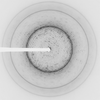
X-ray diffraction data for the Crystal structure of a glucose-1-phosphate thymidylyltransferase from Burkholderia phymatum bound to 2'-deoxy-thymidine-B-L-rhamnose

First author:
Seattle Structural Genomics Center for Infectious Disease (SSGCID)
Resolution: 2.10 Å
R/Rfree: 0.18/0.23
Resolution: 2.10 Å
R/Rfree: 0.18/0.23
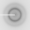
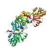
X-ray diffraction data for the Crystal Structure of Ribose-phosphate Pyrophosphokinase from Legionella pneumophila with bound AMP, ADP, and Ribose-5-Phosphate
First author:
Seattle Structural Genomics Center for Infectious Disease (SSGCID)
Resolution: 1.75 Å
R/Rfree: 0.15/0.18
Resolution: 1.75 Å
R/Rfree: 0.15/0.18
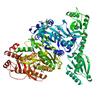
X-ray diffraction data for the Crystal structure of prolyl-tRNA synthetase from Naegleria fowleri in complex with proline and adenosine monophophsphate (AMP)

First author:
Seattle Structural Genomics Center for Infectious Disease (SSGCID)
Resolution: 2.00 Å
R/Rfree: 0.15/0.19
Resolution: 2.00 Å
R/Rfree: 0.15/0.19
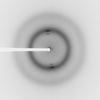
X-ray diffraction data for the Crystal structure of N-myristoyl transferase (NMT) G386E mutant from Plasmodium vivax in complex with inhibitor IMP-1002
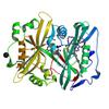
First author:
Seattle Structural Genomics Center for Infectious Disease (SSGCID)
Resolution: 1.55 Å
R/Rfree: 0.15/0.18
Resolution: 1.55 Å
R/Rfree: 0.15/0.18
X-ray diffraction data for the Crystal structure of N-myristoyl transferase (NMT) G386E mutant from Plasmodium vivax in complex with inhibitor IMP-0366
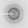
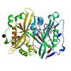
First author:
Seattle Structural Genomics Center for Infectious Disease (SSGCID)
Resolution: 1.55 Å
R/Rfree: 0.15/0.17
Resolution: 1.55 Å
R/Rfree: 0.15/0.17