Diffraction project datasets 3u04
- Method: Molecular Replacement
- Resolution: 1.7 Å
- Space group: C 1 2 1
PDB website for 3U04
PDB validation report for 3U04
doi:10.18430/M33U04
Project details
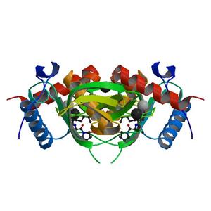
| Title | Crystal structure of peptide deformylase from ehrlichia chaffeensis in complex with actinonin |
| Authors | Seattle Structural Genomics Center for Infectious Disease (SSGCID), Abendroth, J., Clifton, M.C., Edwards, T.E., Staker, B.L. |
| R / Rfree | 0.17 / 0.20 |
| Unit cell edges [Å] | 83.89 x 33.02 x 68.14 |
| Unit cell angles [°] | 90.0, 91.2, 90.0 |
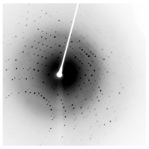
Dataset 225749g7_x####.img details
| Number of frames | 720 (1 - 720) |
| Distance [mm] | 50.0 |
| Oscillation width [°] | 0.50 |
| Omega [°] | -80.0 |
| Wavelength [Å] | 1.54178 |
| Equipment | HOME_SOURCE at HOME_SOURCE (Home Source) |
Dataset 225749g7_y####.img details
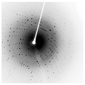
| Number of frames | 720 (1 - 720) |
| Distance [mm] | 50.0 |
| Oscillation width [°] | 0.50 |
| Omega [°] | -80.0 |
| Kappa / Chi [°] | 15.0 |
| Wavelength [Å] | 1.54178 |
| Equipment | HOME_SOURCE at HOME_SOURCE (Home Source) |
Dataset 225749g7_z####.img details
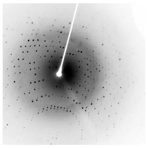
| Number of frames | 720 (1 - 720) |
| Distance [mm] | 50.0 |
| Oscillation width [°] | 0.50 |
| Omega [°] | -80.0 |
| Kappa / Chi [°] | 30.0 |
| Wavelength [Å] | 1.54178 |
| Equipment | HOME_SOURCE at HOME_SOURCE (Home Source) |