1764 results
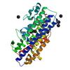
X-ray diffraction data for the Crystal structure of a putative iron-regulated protein A precursor (BDI_2603) from Parabacteroides distasonis ATCC 8503 at 2.30 A resolution
First author:
JOINT CENTER FOR STRUCTURAL GENOMICS (JCSG)
Resolution: 2.30 Å
R/Rfree: 0.16/0.19
Resolution: 2.30 Å
R/Rfree: 0.16/0.19
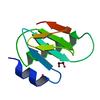
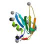
X-ray diffraction data for the Crystal structure of a protein with alpha-lytic protease prodomain-like fold (BDI_0842) from Parabacteroides distasonis ATCC 8503 at 1.40 A resolution
First author:
JOINT CENTER FOR STRUCTURAL GENOMICS (JCSG)
Resolution: 1.40 Å
R/Rfree: 0.13/0.17
Resolution: 1.40 Å
R/Rfree: 0.13/0.17
X-ray diffraction data for the Crystal structure of S-adenosylmethionine decarboxylase proenzyme (TM0655) from THERMOTOGA MARITIMA at 1.2 A resolution
First author:
Joint Center for Structural Genomics (JCSG)
Resolution: 1.20 Å
Resolution: 1.20 Å
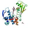
X-ray diffraction data for the Crystal structure of Putative metal-dependent phosphohydrolase (YP_926882.1) from Shewanella amazonensis SB2B at 2.06 A resolution
First author:
Joint Center for Structural Genomics (JCSG)
Resolution: 2.06 Å
R/Rfree: 0.19/0.24
Resolution: 2.06 Å
R/Rfree: 0.19/0.24
X-ray diffraction data for the 1.45 Angstrom Resolution Crystal Structure of PDZ domain of Carboxy-Terminal Protease from Vibrio cholerae in Complex with Peptide.
X-ray diffraction data for the 2.6 Angstrom Resolution Crystal Structure of Stage II Sporulation Protein D (SpoIID) from Clostridium difficile in Complex with Triacetylchitotriose
X-ray diffraction data for the 1.93 Angstrom resolution crystal structure of a pullulanase-specific type II secretion system integral cytoplasmic membrane protein GspL (C-terminal fragment; residues 309-397) from Klebsiella pneumoniae subsp. pneumoniae NTUH-K2044
First author:
A.S. Halavaty
Uniprot: A0A060VDE2
Gene name: pulL
Resolution: 1.93 Å
R/Rfree: 0.23/0.28
Uniprot: A0A060VDE2
Gene name: pulL
Resolution: 1.93 Å
R/Rfree: 0.23/0.28
X-ray diffraction data for the 2.2 Angstrom Resolution Crystal Structure of P-Hydroxybenzoate Hydroxylase from Pseudomonas putida in Complex with FAD.

X-ray diffraction data for the 2.75 Angstrom Resolution Crystal Structure of UDP-N-acetylglucosamine 1-carboxyvinyltransferase from Pseudomonas putida in Complex with Uridine-diphosphate-2(n-acetylglucosaminyl) butyric acid, (2R)-2-(phosphonooxy)propanoic acid and Magnesium
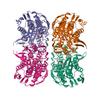
X-ray diffraction data for the The crystal structure of the DPS2 from DEINOCOCCUS RADIODURANS to 1.83A resolution (sequentially soaked in CaCl2 [5mM] for 20 min, then in Ammonium iron(II) sulfate [10mM] for 2h).
First author:
M.G. Cuypers
Resolution: 1.83 Å
R/Rfree: 0.17/0.21
Resolution: 1.83 Å
R/Rfree: 0.17/0.21