6929 results
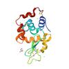
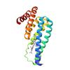
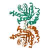
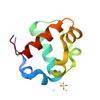
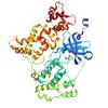
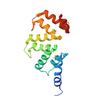
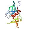
X-ray diffraction data for the Crystal Structure of Histone-lysine N-methyltransferase from Leishmania major in complex with S-ADENOSYLMETHIONINE
First author:
Seattle Structural Genomics Center for Infectious Disease (SSGCID)
Uniprot: Q4QIU2
Resolution: 1.93 Å
R/Rfree: 0.18/0.23
Uniprot: Q4QIU2
Resolution: 1.93 Å
R/Rfree: 0.18/0.23
X-ray diffraction data for the 9p5a
First author:
Fernando Ribeiro
X-ray diffraction data for the 9p5d
First author:
Fernando Ribeiro
X-ray diffraction data for the 9p5e
First author:
Fernando Ribeiro
X-ray diffraction data for the 9p5g
First author:
Fernando Ribeiro
X-ray diffraction data for the 9p5f
First author:
Fernando Ribeiro
X-ray diffraction data for the 9p56
First author:
Fernando Ribeiro
X-ray diffraction data for the 9p59
First author:
Fernando Ribeiro
X-ray diffraction data for the 9p57
First author:
Fernando Ribeiro
X-ray diffraction data for the 9p58
First author:
Fernando Ribeiro
X-ray diffraction data for the LemaA_00628_a_B2-ADP-AMP_37ap
First author:
Diffraction Data Upload SSGCID
X-ray diffraction data for the LemaA_00628_a_B2-ADP-AMP_37as
First author:
Diffraction Data Upload SSGCID
X-ray diffraction data for the 11af
First author:
KARTHIK SELVAM
X-ray diffraction data for the 9p5b
First author:
Fernando Ribeiro
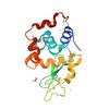
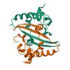
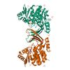
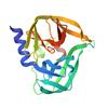
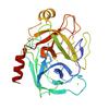
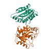
X-ray diffraction data for the Crystal structure of a Phosphoribosylaminoimidazole carboxylase from Burkholderia xenovorans (Apo, Orthorhombic P form)
First author:
Seattle Structural Genomics Center for Infectious Disease (SSGCID)
Uniprot: Q13UJ9
Resolution: 2.05 Å
R/Rfree: 0.18/0.23
Uniprot: Q13UJ9
Resolution: 2.05 Å
R/Rfree: 0.18/0.23
X-ray diffraction data for the D-GlcNAc-bound structure of Vibrio vulnificus putative carbohydrate binding module and split domain
First author:
B.P. Soares
Resolution: 2.28 Å
R/Rfree: 0.19/0.22
Resolution: 2.28 Å
R/Rfree: 0.19/0.22
X-ray diffraction data for the Structure of de novo designed doxorubicin binding protein with doxorubicin bound
First author:
M. Horst
Resolution: 1.56 Å
R/Rfree: 0.22/0.26
Resolution: 1.56 Å
R/Rfree: 0.22/0.26
X-ray diffraction data for the Crystal Structure of cGMP-dependent protein kinase from Plasmodium vivax in complex with inhibitor RUBP-60
First author:
Seattle Structural Genomics Center for Infectious Disease (SSGCID) Seattle Structural Genomics Center for Infectious Disease
Uniprot: A5K0N4
Resolution: 2.90 Å
R/Rfree: 0.22/0.27
Uniprot: A5K0N4
Resolution: 2.90 Å
R/Rfree: 0.22/0.27
X-ray diffraction data for the Crystal structure of hen egg white lysozyme at 100 Kelvin with PEG 6000

X-ray diffraction data for the Crystal structure of hen egg white lysozyme at 100 Kelvin with PEG 400 (Triplicate)

X-ray diffraction data for the Crystal structure of hen egg white lysozyme at 100 Kelvin with PEG 6000 (Duplicate)
X-ray diffraction data for the Crystal structure of hen egg white lysozyme at 100 Kelvin with PEG 400 (Duplicate)
X-ray diffraction data for the Crystal structure of hen egg white lysozyme at 100 Kelvin with PEG 6000 (Triplicate)

X-ray diffraction data for the Crystal structure of hen egg white lysozyme at 100 Kelvin with Silicone Oil (Triplicate)
X-ray diffraction data for the Crystal structure of hen egg white lysozyme at 100 Kelvin with Vaseline (Duplicate)
X-ray diffraction data for the Crystal structure of hen egg white lysozyme at 100 Kelvin with Vaseline
X-ray diffraction data for the Crystal structure of hen egg white lysozyme at 100 Kelvin with Silicone Oil
X-ray diffraction data for the Crystal structure of hen egg white lysozyme at 100 Kelvin with Silicone Oil (Duplicate)
X-ray diffraction data for the Crystal structure of hen egg white lysozyme at 300 Kelvin with Apiezon N (Triplicate)
X-ray diffraction data for the Crystal structure of hen egg white lysozyme at 100 Kelvin with Vaseline (Triplicate)

X-ray diffraction data for the Crystal structure of hen egg white lysozyme at 300 Kelvin with Vaseline
X-ray diffraction data for the Crystal structure of hen egg white lysozyme at 300 Kelvin with Apiezon N
X-ray diffraction data for the Crystal structure of hen egg white lysozyme at 300 Kelvin with Vaseline (Duplicate)

X-ray diffraction data for the Crystal structure of hen egg white lysozyme at 300 Kelvin with Vaseline (Triplicate)

X-ray diffraction data for the Crystal structure of hen egg white lysozyme at 300 Kelvin with Apiezon T (Triplicate)

X-ray diffraction data for the Crystal structure of hen egg white lysozyme at 300 Kelvin with Apiezon T (Duplicate)

X-ray diffraction data for the Crystal structure of hen egg white lysozyme at 300 Kelvin with Apiezon T
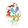
X-ray diffraction data for the Crystal structure of a large ribosomal subunit protein bL17 from Bordetella pertussis
First author:
Seattle Structural Genomics Center for Infectious Disease (SSGCID)
Uniprot: Q7VTA6
Resolution: 1.65 Å
R/Rfree: 0.19/0.22
Uniprot: Q7VTA6
Resolution: 1.65 Å
R/Rfree: 0.19/0.22
X-ray diffraction data for the psaea_00612_a_b3-36lw
First author:
Diffraction Data Upload SSGCID
X-ray diffraction data for the The dose series study of NcAA9D: wedge 20-38
First author:
Sam Miller
X-ray diffraction data for the The dose series study of NcAA9D: wedge 1–19
First author:
Sam Miller
X-ray diffraction data for the Crystal structure of hen egg white lysozyme at 100 Kelvin with Apiezon N

X-ray diffraction data for the Crystal structure of hen egg white lysozyme at 100 Kelvin with Apiezon N (Duplicate)
X-ray diffraction data for the Crystal structure of hen egg white lysozyme at 100 Kelvin with Lard (Triplicate)

X-ray diffraction data for the Crystal structure of hen egg white lysozyme at 100 Kelvin with Apiezon N (Triplicate)

X-ray diffraction data for the Crystal structure of Phosphoribosylaminoimidazole carboxylase from Burkholderia xenovorans (Apo)
First author:
Seattle Structural Genomics Center for Infectious Disease (SSGCID)
Uniprot: Q13UJ9
Resolution: 2.39 Å
R/Rfree: 0.23/0.28
Uniprot: Q13UJ9
Resolution: 2.39 Å
R/Rfree: 0.23/0.28
X-ray diffraction data for the Crystal structure of the cysteine-free anti-UTag intrabody
First author:
S.H. Chen
Resolution: 1.45 Å
R/Rfree: 0.17/0.21
Resolution: 1.45 Å
R/Rfree: 0.17/0.21
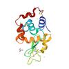
X-ray diffraction data for the Crystal structure of human adenosine kinase (ADK) in complex with inhibitor BKI-1676
First author:
Seattle Structural Genomics Center for Infectious Disease (SSGCID)
Uniprot: P55263
Resolution: 2.81 Å
R/Rfree: 0.24/0.27
Uniprot: P55263
Resolution: 2.81 Å
R/Rfree: 0.24/0.27
X-ray diffraction data for the Crystal structure of human adenosine kinase (ADK) in complex with inhibitor BKI-1817
First author:
Seattle Structural Genomics Center for Infectious Disease (SSGCID)
Uniprot: P55263
Resolution: 2.51 Å
R/Rfree: 0.21/0.25
Uniprot: P55263
Resolution: 2.51 Å
R/Rfree: 0.21/0.25
X-ray diffraction data for the Crystal structure of human adenosine kinase (ADK) in complex with inhibitor BKI-1553

First author:
Seattle Structural Genomics Center for Infectious Disease (SSGCID)
Uniprot: P55263
Resolution: 2.41 Å
R/Rfree: 0.22/0.26
Uniprot: P55263
Resolution: 2.41 Å
R/Rfree: 0.22/0.26
X-ray diffraction data for the Transcription factor
X-ray diffraction data for the Doc and Phd complex from Enterococcus faecalis
X-ray diffraction data for the Antitoxin Phd
X-ray diffraction data for the Crystal structure of Enterovirus A71 2A protease mutant C110A containing VP1-2A junction in the active site determined via sulfur phasing

X-ray diffraction data for the Crystal structure of a SnoaL-like domain-containing protein from Mycobacterium ulcerans (Zinc Bound)
First author:
Seattle Structural Genomics Center for Infectious Disease (SSGCID) Seattle Structural Genomics Center for Infectious Disease
Uniprot: A0PR67
Resolution: 1.31 Å
R/Rfree: 0.15/0.17
Uniprot: A0PR67
Resolution: 1.31 Å
R/Rfree: 0.15/0.17
X-ray diffraction data for the Sterile Alpha Motif of Human Translocation ETS Leukemia, Non-Polymer Crystal Form
X-ray diffraction data for the 1TEL, non-compressed, double-helical crystal form
X-ray diffraction data for the Crystal Structure of serine/threonine-protein kinase (AEK1) from Trypanosoma cruzi in complex with AMP
First author:
Seattle Structural Genomics Center for Infectious Disease (SSGCID)
Uniprot: Q4E2L0
Resolution: 2.83 Å
R/Rfree: 0.23/0.28
Uniprot: Q4E2L0
Resolution: 2.83 Å
R/Rfree: 0.23/0.28
X-ray diffraction data for the Crystal Structure of serine/threonine-protein kinase (AEK1) from Trypanosoma cruzi in complex with ATP
First author:
Seattle Structural Genomics Center for Infectious Disease (SSGCID)
Uniprot: Q4E2L0
Resolution: 2.68 Å
R/Rfree: 0.23/0.28
Uniprot: Q4E2L0
Resolution: 2.68 Å
R/Rfree: 0.23/0.28
X-ray diffraction data for the Crystal Structure of serine/threonine-protein kinase (AEK1) from Trypanosoma cruzi in complex with LMS
First author:
Seattle Structural Genomics Center for Infectious Disease (SSGCID)
Uniprot: Q4E2L0
Resolution: 2.82 Å
R/Rfree: 0.24/0.28
Uniprot: Q4E2L0
Resolution: 2.82 Å
R/Rfree: 0.24/0.28
X-ray diffraction data for the Crystal Structure of Superoxide dismutase from Cryptosporidium parvum
First author:
Seattle Structural Genomics Center for Infectious Disease (SSGCID)
Uniprot: A3FQJ9
Resolution: 2.01 Å
R/Rfree: 0.15/0.20
Uniprot: A3FQJ9
Resolution: 2.01 Å
R/Rfree: 0.15/0.20
X-ray diffraction data for the Structure of de novo designed salen-binding enzyme

First author:
I. Bakanas
Resolution: 1.99 Å
R/Rfree: 0.21/0.26
Resolution: 1.99 Å
R/Rfree: 0.21/0.26
X-ray diffraction data for the Structure of de novo designed salen-binding enzyme with salen bound
First author:
I. Bakanas
Resolution: 1.66 Å
R/Rfree: 0.23/0.26
Resolution: 1.66 Å
R/Rfree: 0.23/0.26
X-ray diffraction data for the Dimeric C-terminal domain of Nucleocapsid protein of SARS-CoV-2.
X-ray diffraction data for the Crystal Structure of L-erythrulose-1-phosphate isomerase from Brucella melitensis in complex with SN-GLYCEROL-1-PHOSPHATE
First author:
Seattle Structural Genomics Center for Infectious Disease (SSGCID)
Uniprot: Q2YIQ6
Resolution: 1.95 Å
R/Rfree: 0.16/0.22
Uniprot: Q2YIQ6
Resolution: 1.95 Å
R/Rfree: 0.16/0.22
X-ray diffraction data for the Crystal Structure of a Ribokinase from Brucella suis in complex ATP (P21 form)
First author:
Seattle Structural Genomics Center for Infectious Disease (SSGCID)
Uniprot: A0A0H3GDY9
Resolution: 2.60 Å
R/Rfree: 0.20/0.23
Uniprot: A0A0H3GDY9
Resolution: 2.60 Å
R/Rfree: 0.20/0.23
X-ray diffraction data for the Sterile Alpha Motif Domain of Human Translocation ETS Leukemia, Non-Polymer Crystal Form
X-ray diffraction data for the Crystal structure of Enterovirus D68 3C protease determined via sulfur phasing

X-ray diffraction data for the Crystal structure of calcium-dependent protein kinase 1 (CDPK1) from Cryptosporidium parvum in complex with inhibitor WIN-1-158.
First author:
Seattle Structural Genomics Center for Infectious Disease (SSGCID) Seattle Structural Genomics Center for Infectious Disease
Uniprot: A3FQ16
Resolution: 2.21 Å
R/Rfree: 0.20/0.24
Uniprot: A3FQ16
Resolution: 2.21 Å
R/Rfree: 0.20/0.24
X-ray diffraction data for the Crystal structure of calcium-dependent protein kinase 1 (CDPK1) from Cryptosporidium parvum in complex with inhibitor BKI-1862.
First author:
Seattle Structural Genomics Center for Infectious Disease (SSGCID) Seattle Structural Genomics Center for Infectious Disease
Uniprot: A3FQ16
Resolution: 2.52 Å
R/Rfree: 0.21/0.26
Uniprot: A3FQ16
Resolution: 2.52 Å
R/Rfree: 0.21/0.26
X-ray diffraction data for the Crystal structure of calcium-dependent protein kinase 1 (CDPK1) from Cryptosporidium parvum in complex with inhibitor WIN-1-159.
First author:
Seattle Structural Genomics Center for Infectious Disease (SSGCID) Seattle Structural Genomics Center for Infectious Disease
Uniprot: A3FQ16
Resolution: 2.30 Å
R/Rfree: 0.21/0.25
Uniprot: A3FQ16
Resolution: 2.30 Å
R/Rfree: 0.21/0.25
X-ray diffraction data for the Crystal structure of calcium-dependent protein kinase 1 (CDPK1) from Cryptosporidium parvum in complex with inhibitor BKI-1708.

First author:
Seattle Structural Genomics Center for Infectious Disease (SSGCID) Seattle Structural Genomics Center for Infectious Disease
Uniprot: A3FQ16
Resolution: 2.20 Å
R/Rfree: 0.20/0.26
Uniprot: A3FQ16
Resolution: 2.20 Å
R/Rfree: 0.20/0.26
X-ray diffraction data for the Crystal structure of calcium-dependent protein kinase 1 (CDPK1) from Cryptosporidium parvum in complex with inhibitor BKI-1932.
First author:
Seattle Structural Genomics Center for Infectious Disease (SSGCID) Seattle Structural Genomics Center for Infectious Disease
Uniprot: A3FQ16
Resolution: 1.84 Å
R/Rfree: 0.19/0.21
Uniprot: A3FQ16
Resolution: 1.84 Å
R/Rfree: 0.19/0.21
X-ray diffraction data for the Crystal structure of calcium-dependent protein kinase 1 (CDPK1) from Cryptosporidium parvum in complex with inhibitor WIN-III-6.
First author:
Seattle Structural Genomics Center for Infectious Disease (SSGCID) Seattle Structural Genomics Center for Infectious Disease
Uniprot: A3FQ16
Resolution: 1.94 Å
R/Rfree: 0.19/0.23
Uniprot: A3FQ16
Resolution: 1.94 Å
R/Rfree: 0.19/0.23
X-ray diffraction data for the Crystal structure of Zika virus NS2B-NS3 protease determined via sulfur phasing


X-ray diffraction data for the Crystal structure of trypsin at 100 Kelvin with benzamidine (triplicate)
X-ray diffraction data for the KEAP1 complexed to cyclic peptide 33
X-ray diffraction data for the KEAP1 complexed to cyclic peptide 30
X-ray diffraction data for the KEAP1 complexed to cyclic peptide 34
X-ray diffraction data for the KOD-H4 DNA polymerase mutant in a binary complex with DNA:DNA containing two AtNA nucleotides
X-ray diffraction data for the Crystal Structure of ribosomal large subunit pseudouridine synthase D from Neisseria gonorrhoea
First author:
Seattle Structural Genomics Center for Infectious Disease (SSGCID)
Uniprot: B4RJW3
Resolution: 1.91 Å
R/Rfree: 0.17/0.19
Uniprot: B4RJW3
Resolution: 1.91 Å
R/Rfree: 0.17/0.19
X-ray diffraction data for the Crystal structure of Cysteinyl-tRNA synthetase (CysRS) from Plasmodium falciparum in complex with ADP (long soak)
First author:
Seattle Structural Genomics Center for Infectious Disease (SSGCID)
Uniprot: Q8IJP3
Resolution: 2.45 Å
R/Rfree: 0.19/0.22
Uniprot: Q8IJP3
Resolution: 2.45 Å
R/Rfree: 0.19/0.22
X-ray diffraction data for the Crystal Structure of ATP phosphoribosyltransferase from Bordetella pertussis

First author:
Seattle Structural Genomics Center for Infectious Disease (SSGCID)
Uniprot: Q7VSZ2
Resolution: 1.55 Å
R/Rfree: 0.19/0.22
Uniprot: Q7VSZ2
Resolution: 1.55 Å
R/Rfree: 0.19/0.22
X-ray diffraction data for the Crystal structure of Coxsackievirus A16 (G-10) 2A protease determined via sulfur phasing

X-ray diffraction data for the Crystal structure of Ami1 from M. tuberculosis in complex with a tetrazole compound
X-ray diffraction data for the Crystal structure of Borneoldehydrogenase ancestor N39
First author:
C.P.O. Helmer
Resolution: 1.85 Å
R/Rfree: 0.17/0.21
Resolution: 1.85 Å
R/Rfree: 0.17/0.21
X-ray diffraction data for the Helicobacter pylori cysteine rich protein B (hcpB)
X-ray diffraction data for the Crystallizing extracellular protein reveals paths for silver mineralization and recovery
X-ray diffraction data for the Crystal structure of PHICD111_20024_EAD.
X-ray diffraction data for the Crystal structure of C. merolae LAMMER-like dual specificity kinase (CmLIK) kinase domain
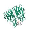
X-ray diffraction data for the Neutralizing monoclonal antibody Fab fragment for human leptin

First author:
D.R. Tomchick
Resolution: 3.30 Å
R/Rfree: 0.27/0.32
Resolution: 3.30 Å
R/Rfree: 0.27/0.32
X-ray diffraction data for the Crystal Structure of L-erythrulose-1-phosphate isomerase from Brucella melitensis (P1 form)
First author:
Seattle Structural Genomics Center for Infectious Disease (SSGCID)
Uniprot: Q2YIQ6
Resolution: 1.60 Å
R/Rfree: 0.16/0.18
Uniprot: Q2YIQ6
Resolution: 1.60 Å
R/Rfree: 0.16/0.18
X-ray diffraction data for the Crystal Structure of L-erythrulose-1-phosphate isomerase from Brucella melitensis (P21 form)
First author:
Seattle Structural Genomics Center for Infectious Disease (SSGCID)
Uniprot: Q2YIQ6
Resolution: 2.15 Å
R/Rfree: 0.18/0.22
Uniprot: Q2YIQ6
Resolution: 2.15 Å
R/Rfree: 0.18/0.22
X-ray diffraction data for the Drosophila melanogaster setdb1-tuor domain
X-ray diffraction data for the Co-crystal structure of human CARM1 in complex with MT556 inhibitor
X-ray diffraction data for the Crystal structure of the WDR domain of human DCAF1 in complex with OICR-6766

X-ray diffraction data for the Co-crystal structure of 53BP1 tandem Tudor domains in complex with UNC8531
X-ray diffraction data for the Crystal structure of Capsular polysaccharide biosynthesis protein from Bordetella pertussis in complex with NAD and uridine-diphosphate-n-acetylgalactosamine (cocrystallization)
First author:
Seattle Structural Genomics Center for Infectious Disease (SSGCID)
Uniprot: Q7TTK0
Resolution: 1.70 Å
R/Rfree: 0.15/0.18
Uniprot: Q7TTK0
Resolution: 1.70 Å
R/Rfree: 0.15/0.18
X-ray diffraction data for the Crystal Structure of serine/threonine-protein kinase (AEK1) T376D, S395D Mutant from Trypanosoma brucei (AMP-PNP)
First author:
Seattle Structural Genomics Center for Infectious Disease (SSGCID)
Uniprot: Q582V7
Resolution: 2.15 Å
R/Rfree: 0.22/0.25
Uniprot: Q582V7
Resolution: 2.15 Å
R/Rfree: 0.22/0.25
X-ray diffraction data for the Crystal structure of bacterial extracellular solute-binding protein from Bordetella bronchiseptica RB50

First author:
C. Chang
Resolution: 1.55 Å
R/Rfree: 0.17/0.19
Resolution: 1.55 Å
R/Rfree: 0.17/0.19