6862 results
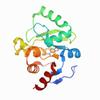
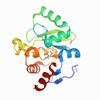
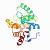
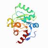
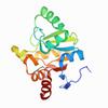
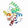
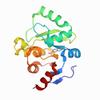
X-ray diffraction data for the Crystal structure of SARS-CoV-2 NSP3 macrodomain in complex with ZINC000039281982
First author:
G.J. Correy
Resolution: 1.05 Å
R/Rfree: 0.20/0.22
Resolution: 1.05 Å
R/Rfree: 0.20/0.22
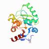
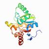
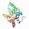
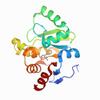
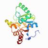
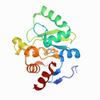
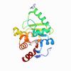
X-ray diffraction data for the Crystal structure of SARS-CoV-2 NSP3 macrodomain in complex with ZINC000008652361

First author:
G.J. Correy
Resolution: 1.00 Å
R/Rfree: 0.16/0.17
Resolution: 1.00 Å
R/Rfree: 0.16/0.17
X-ray diffraction data for the Crystal structure of SARS-CoV-2 NSP3 macrodomain in complex with ZINC000000039810
First author:
G.J. Correy
Resolution: 1.00 Å
R/Rfree: 0.15/0.16
Resolution: 1.00 Å
R/Rfree: 0.15/0.16
X-ray diffraction data for the Crystal structure of SARS-CoV-2 NSP3 macrodomain in complex with ZINC000002582714
First author:
G.J. Correy
Resolution: 1.00 Å
R/Rfree: 0.14/0.16
Resolution: 1.00 Å
R/Rfree: 0.14/0.16
X-ray diffraction data for the Crystal structure of SARS-CoV-2 NSP3 macrodomain in complex with ZINC000004218283

First author:
G.J. Correy
Resolution: 1.02 Å
R/Rfree: 0.17/0.18
Resolution: 1.02 Å
R/Rfree: 0.17/0.18
X-ray diffraction data for the Crystal structure of SARS-CoV-2 NSP3 macrodomain in complex with ZINC000004787230
First author:
G.J. Correy
Resolution: 1.04 Å
R/Rfree: 0.17/0.19
Resolution: 1.04 Å
R/Rfree: 0.17/0.19
X-ray diffraction data for the Crystal structure of SARS-CoV-2 NSP3 macrodomain in complex with ZINC000000388262
First author:
G.J. Correy
Resolution: 1.00 Å
R/Rfree: 0.16/0.18
Resolution: 1.00 Å
R/Rfree: 0.16/0.18
X-ray diffraction data for the Crystal structure of SARS-CoV-2 NSP3 macrodomain in complex with ZINC000000337835

First author:
G.J. Correy
Resolution: 1.00 Å
R/Rfree: 0.16/0.18
Resolution: 1.00 Å
R/Rfree: 0.16/0.18
X-ray diffraction data for the Crystal structure of SARS-CoV-2 NSP3 macrodomain in complex with ZINC000000340465

First author:
G.J. Correy
Resolution: 1.03 Å
R/Rfree: 0.16/0.18
Resolution: 1.03 Å
R/Rfree: 0.16/0.18
X-ray diffraction data for the Crystal structure of SARS-CoV-2 NSP3 macrodomain in complex with ZINC000000002055
First author:
G.J. Correy
Resolution: 1.00 Å
R/Rfree: 0.15/0.16
Resolution: 1.00 Å
R/Rfree: 0.15/0.16
X-ray diffraction data for the Crystal structure of SARS-CoV-2 NSP3 macrodomain in complex with ZINC000000332673
First author:
G.J. Correy
Resolution: 1.00 Å
R/Rfree: 0.15/0.17
Resolution: 1.00 Å
R/Rfree: 0.15/0.17
X-ray diffraction data for the Crystal structure of SARS-CoV-2 NSP3 macrodomain in complex with ZINC000006691828
First author:
G.J. Correy
Resolution: 1.00 Å
R/Rfree: 0.15/0.17
Resolution: 1.00 Å
R/Rfree: 0.15/0.17
X-ray diffraction data for the Crystal structure of SARS-CoV-2 NSP3 macrodomain in complex with ZINC000000158650
First author:
G.J. Correy
Resolution: 1.00 Å
R/Rfree: 0.17/0.19
Resolution: 1.00 Å
R/Rfree: 0.17/0.19
X-ray diffraction data for the Crystal structure of SARS-CoV-2 NSP3 macrodomain in complex with ZINC000000158490
First author:
G.J. Correy
Resolution: 1.00 Å
R/Rfree: 0.16/0.17
Resolution: 1.00 Å
R/Rfree: 0.16/0.17
X-ray diffraction data for the Crystal structure of SARS-CoV-2 NSP3 macrodomain in complex with ZINC000002560357
First author:
G.J. Correy
Resolution: 1.02 Å
R/Rfree: 0.16/0.17
Resolution: 1.02 Å
R/Rfree: 0.16/0.17
X-ray diffraction data for the Crystal structure of SARS-CoV-2 NSP3 macrodomain in complex with ZINC000000000226

First author:
G.J. Correy
Resolution: 1.03 Å
R/Rfree: 0.17/0.19
Resolution: 1.03 Å
R/Rfree: 0.17/0.19
X-ray diffraction data for the Crystal structure of SARS-CoV-2 NSP3 macrodomain in complex with ZINC000000064576
First author:
G.J. Correy
Resolution: 1.00 Å
R/Rfree: 0.17/0.18
Resolution: 1.00 Å
R/Rfree: 0.17/0.18
X-ray diffraction data for the Crystal structure of SARS-CoV-2 NSP3 macrodomain in complex with ZINC000000001099

First author:
G.J. Correy
Resolution: 1.02 Å
R/Rfree: 0.17/0.18
Resolution: 1.02 Å
R/Rfree: 0.17/0.18
X-ray diffraction data for the Crystal structure of SARS-CoV-2 NSP3 macrodomain in complex with ZINC000000331715
First author:
G.J. Correy
Resolution: 1.00 Å
R/Rfree: 0.18/0.19
Resolution: 1.00 Å
R/Rfree: 0.18/0.19
X-ray diffraction data for the Crystal structure of SARS-CoV-2 NSP3 macrodomain in complex with ZINC000000034687
First author:
G.J. Correy
Resolution: 1.00 Å
R/Rfree: 0.17/0.18
Resolution: 1.00 Å
R/Rfree: 0.17/0.18
X-ray diffraction data for the Crystal structure of SARS-CoV-2 NSP3 macrodomain in complex with ZINC000084843283
First author:
G.J. Correy
Resolution: 0.94 Å
R/Rfree: 0.17/0.18
Resolution: 0.94 Å
R/Rfree: 0.17/0.18
X-ray diffraction data for the Crystal structure of SARS-CoV-2 NSP3 macrodomain in complex with ZINC000000265642
First author:
G.J. Correy
Resolution: 0.98 Å
R/Rfree: 0.17/0.18
Resolution: 0.98 Å
R/Rfree: 0.17/0.18
X-ray diffraction data for the Crystal structure of SARS-CoV-2 NSP3 macrodomain in complex with ZINC000400552187_N3
First author:
G.J. Correy
Resolution: 1.00 Å
R/Rfree: 0.16/0.18
Resolution: 1.00 Å
R/Rfree: 0.16/0.18
X-ray diffraction data for the Crystal structure of SARS-CoV-2 NSP3 macrodomain in complex with ZINC000082473428_N3
First author:
G.J. Correy
Resolution: 1.00 Å
R/Rfree: 0.16/0.17
Resolution: 1.00 Å
R/Rfree: 0.16/0.17
X-ray diffraction data for the Crystal structure of SARS-CoV-2 NSP3 macrodomain in complex with ZINC000736709772
First author:
G.J. Correy
Resolution: 1.00 Å
R/Rfree: 0.16/0.17
Resolution: 1.00 Å
R/Rfree: 0.16/0.17