6912 results
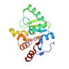
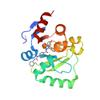
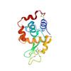
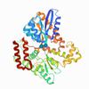
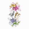
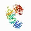
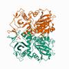
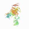
X-ray diffraction data for the PanDDA analysis group deposition -- Crystal structure of SARS-CoV-2 NSP3 macrodomain in complex with FRESH00014134848 - (R) isomer
First author:
G.J. Correy
Resolution: 1.05 Å
R/Rfree: 0.15/0.16
Resolution: 1.05 Å
R/Rfree: 0.15/0.16
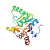
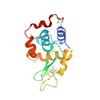
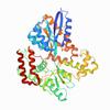
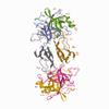
X-ray diffraction data for the PanDDA analysis group deposition -- Crystal structure of SARS-CoV-2 NSP3 macrodomain in complex with REAL250002155324
First author:
G.J. Correy
Resolution: 1.05 Å
R/Rfree: 0.14/0.16
Resolution: 1.05 Å
R/Rfree: 0.14/0.16
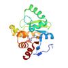
X-ray diffraction data for the PanDDA analysis group deposition -- Crystal structure of SARS-CoV-2 NSP3 macrodomain in complex with FRESH00020289192 - (S) isomer


First author:
G.J. Correy
Resolution: 1.05 Å
R/Rfree: 0.13/0.15
Resolution: 1.05 Å
R/Rfree: 0.13/0.15
X-ray diffraction data for the PanDDA analysis group deposition -- Crystal structure of SARS-CoV-2 NSP3 macrodomain in complex with FRESH00010608284
First author:
G.J. Correy
Resolution: 1.05 Å
R/Rfree: 0.14/0.16
Resolution: 1.05 Å
R/Rfree: 0.14/0.16
X-ray diffraction data for the PanDDA analysis group deposition -- Crystal structure of SARS-CoV-2 NSP3 macrodomain in complex with FRESH00002410346
First author:
G.J. Correy
Resolution: 1.05 Å
R/Rfree: 0.13/0.15
Resolution: 1.05 Å
R/Rfree: 0.13/0.15
X-ray diffraction data for the PanDDA analysis group deposition -- Crystal structure of SARS-CoV-2 NSP3 macrodomain in complex with ZINC000611664196 - (S,S) isomer

First author:
G.J. Correy
Resolution: 1.05 Å
R/Rfree: 0.13/0.14
Resolution: 1.05 Å
R/Rfree: 0.13/0.14
X-ray diffraction data for the PanDDA analysis group deposition -- Crystal structure of SARS-CoV-2 NSP3 macrodomain in complex with REAL250003296134 - (R) isomer

First author:
G.J. Correy
Resolution: 1.05 Å
R/Rfree: 0.14/0.16
Resolution: 1.05 Å
R/Rfree: 0.14/0.16
X-ray diffraction data for the PanDDA analysis group deposition -- Crystal structure of SARS-CoV-2 NSP3 macrodomain in complex with ZINC000893101964
First author:
G.J. Correy
Resolution: 1.05 Å
R/Rfree: 0.13/0.15
Resolution: 1.05 Å
R/Rfree: 0.13/0.15
X-ray diffraction data for the PanDDA analysis group deposition -- Crystal structure of SARS-CoV-2 NSP3 macrodomain in complex with Z4574659604 - (R,R) and (S,S) isomers

First author:
G.J. Correy
Resolution: 1.05 Å
R/Rfree: 0.13/0.15
Resolution: 1.05 Å
R/Rfree: 0.13/0.15
X-ray diffraction data for the PanDDA analysis group deposition -- Crystal structure of SARS-CoV-2 NSP3 macrodomain in complex with Z4718398515 - (R,S) isomer
First author:
G.J. Correy
Resolution: 1.05 Å
R/Rfree: 0.13/0.15
Resolution: 1.05 Å
R/Rfree: 0.13/0.15
X-ray diffraction data for the PanDDA analysis group deposition -- Crystal structure of SARS-CoV-2 NSP3 macrodomain in complex with Z4718398585
First author:
G.J. Correy
Resolution: 1.05 Å
R/Rfree: 0.14/0.16
Resolution: 1.05 Å
R/Rfree: 0.14/0.16
X-ray diffraction data for the PanDDA analysis group deposition -- Crystal structure of SARS-CoV-2 NSP3 macrodomain in complex with Z4718398569
First author:
G.J. Correy
Resolution: 1.05 Å
R/Rfree: 0.14/0.16
Resolution: 1.05 Å
R/Rfree: 0.14/0.16
X-ray diffraction data for the PanDDA analysis group deposition -- Crystal structure of SARS-CoV-2 NSP3 macrodomain in complex with Z4718398531 - (S,S) isomer
First author:
G.J. Correy
Resolution: 1.05 Å
R/Rfree: 0.13/0.14
Resolution: 1.05 Å
R/Rfree: 0.13/0.14
X-ray diffraction data for the PanDDA analysis group deposition -- Crystal structure of SARS-CoV-2 NSP3 macrodomain in complex with Z4718398539 - (R,R) and (S,S) isomers
First author:
G.J. Correy
Resolution: 1.05 Å
R/Rfree: 0.14/0.16
Resolution: 1.05 Å
R/Rfree: 0.14/0.16
X-ray diffraction data for the PanDDA analysis group deposition -- Crystal structure of SARS-CoV-2 NSP3 macrodomain in complex with Z5010894387 - (R,R) and (S,S) isomers

First author:
G.J. Correy
Resolution: 1.05 Å
R/Rfree: 0.13/0.15
Resolution: 1.05 Å
R/Rfree: 0.13/0.15
X-ray diffraction data for the PanDDA analysis group deposition -- Crystal structure of SARS-CoV-2 NSP3 macrodomain in complex with Z5010894404 - (R,R) and (S,S) isomers

First author:
G.J. Correy
Resolution: 1.05 Å
R/Rfree: 0.15/0.17
Resolution: 1.05 Å
R/Rfree: 0.15/0.17
X-ray diffraction data for the PanDDA analysis group deposition -- Crystal structure of SARS-CoV-2 NSP3 macrodomain in complex with Z5010894417 - (R,R) and (S,S) isomers

First author:
G.J. Correy
Resolution: 1.05 Å
R/Rfree: 0.14/0.16
Resolution: 1.05 Å
R/Rfree: 0.14/0.16
X-ray diffraction data for the PanDDA analysis group deposition -- Crystal structure of SARS-CoV-2 NSP3 macrodomain in complex with Z3508769536 - (S) isomer

First author:
G.J. Correy
Resolution: 1.05 Å
R/Rfree: 0.13/0.15
Resolution: 1.05 Å
R/Rfree: 0.13/0.15
X-ray diffraction data for the PanDDA analysis group deposition -- Crystal structure of SARS-CoV-2 NSP3 macrodomain in complex with Z5010894415 - (S) isomer
First author:
G.J. Correy
Resolution: 1.05 Å
R/Rfree: 0.14/0.16
Resolution: 1.05 Å
R/Rfree: 0.14/0.16
X-ray diffraction data for the PanDDA analysis group deposition -- Crystal structure of SARS-CoV-2 NSP3 macrodomain in complex with Z5010903509 - (S,S) isomer
First author:
G.J. Correy
Resolution: 1.05 Å
R/Rfree: 0.13/0.15
Resolution: 1.05 Å
R/Rfree: 0.13/0.15
X-ray diffraction data for the PanDDA analysis group deposition -- Crystal structure of SARS-CoV-2 NSP3 macrodomain in complex with Z4718398580
First author:
G.J. Correy
Resolution: 1.07 Å
R/Rfree: 0.17/0.20
Resolution: 1.07 Å
R/Rfree: 0.17/0.20
X-ray diffraction data for the PanDDA analysis group deposition -- Crystal structure of SARS-CoV-2 NSP3 macrodomain in complex with Z4718398572
First author:
G.J. Correy
Resolution: 1.06 Å
R/Rfree: 0.16/0.18
Resolution: 1.06 Å
R/Rfree: 0.16/0.18
X-ray diffraction data for the PanDDA analysis group deposition -- Crystal structure of SARS-CoV-2 NSP3 macrodomain in complex with ZINC000450476923 - (S,R) isomer


First author:
G.J. Correy
Resolution: 1.01 Å
R/Rfree: 0.14/0.16
Resolution: 1.01 Å
R/Rfree: 0.14/0.16
X-ray diffraction data for the PanDDA analysis group deposition -- Crystal structure of SARS-CoV-2 NSP3 macrodomain in complex with ZINC000579359572 - (R) and (S) isomers

First author:
G.J. Correy
Resolution: 0.97 Å
R/Rfree: 0.14/0.15
Resolution: 0.97 Å
R/Rfree: 0.14/0.15
X-ray diffraction data for the PanDDA analysis group deposition -- Crystal structure of SARS-CoV-2 NSP3 macrodomain in complex with ZINC001472868186
First author:
G.J. Correy
Resolution: 1.03 Å
R/Rfree: 0.15/0.17
Resolution: 1.03 Å
R/Rfree: 0.15/0.17
X-ray diffraction data for the PanDDA analysis group deposition -- Crystal structure of SARS-CoV-2 NSP3 macrodomain in complex with ZINC000681764827
First author:
G.J. Correy
Resolution: 1.05 Å
R/Rfree: 0.13/0.15
Resolution: 1.05 Å
R/Rfree: 0.13/0.15
X-ray diffraction data for the PanDDA analysis group deposition -- Crystal structure of SARS-CoV-2 NSP3 macrodomain in complex with ZINC000827900828
First author:
G.J. Correy
Resolution: 1.05 Å
R/Rfree: 0.14/0.16
Resolution: 1.05 Å
R/Rfree: 0.14/0.16
X-ray diffraction data for the PanDDA analysis group deposition -- Crystal structure of SARS-CoV-2 NSP3 macrodomain in complex with ZINC000222377450
First author:
G.J. Correy
Resolution: 1.05 Å
R/Rfree: 0.16/0.18
Resolution: 1.05 Å
R/Rfree: 0.16/0.18
X-ray diffraction data for the PanDDA analysis group deposition -- Crystal structure of SARS-CoV-2 NSP3 macrodomain in complex with ZINC000043461211
First author:
G.J. Correy
Resolution: 1.05 Å
R/Rfree: 0.12/0.14
Resolution: 1.05 Å
R/Rfree: 0.12/0.14
X-ray diffraction data for the PanDDA analysis group deposition -- Crystal structure of SARS-CoV-2 NSP3 macrodomain in complex with ZINC000118179920
First author:
G.J. Correy
Resolution: 1.05 Å
R/Rfree: 0.16/0.18
Resolution: 1.05 Å
R/Rfree: 0.16/0.18
X-ray diffraction data for the PanDDA analysis group deposition -- Crystal structure of SARS-CoV-2 NSP3 macrodomain in complex with ZINC000893191027 - (S) and (R) isomers
First author:
G.J. Correy
Resolution: 1.05 Å
R/Rfree: 0.14/0.16
Resolution: 1.05 Å
R/Rfree: 0.14/0.16
X-ray diffraction data for the PanDDA analysis group deposition -- Crystal structure of SARS-CoV-2 NSP3 macrodomain in complex with ZINC000285507655 - (R) and (S) isomers

First author:
G.J. Correy
Resolution: 1.05 Å
R/Rfree: 0.14/0.15
Resolution: 1.05 Å
R/Rfree: 0.14/0.15
X-ray diffraction data for the PanDDA analysis group deposition -- Crystal structure of SARS-CoV-2 NSP3 macrodomain in complex with ZINC000292637864 - (R) and (S) isomers

First author:
G.J. Correy
Resolution: 1.05 Å
R/Rfree: 0.14/0.15
Resolution: 1.05 Å
R/Rfree: 0.14/0.15
X-ray diffraction data for the PanDDA analysis group deposition -- Crystal structure of SARS-CoV-2 NSP3 macrodomain in complex with ZINC000559260078
First author:
G.J. Correy
Resolution: 1.05 Å
R/Rfree: 0.13/0.15
Resolution: 1.05 Å
R/Rfree: 0.13/0.15
X-ray diffraction data for the PanDDA analysis group deposition -- Crystal structure of SARS-CoV-2 NSP3 macrodomain in complex with ZINC000110510893
First author:
G.J. Correy
Resolution: 1.05 Å
R/Rfree: 0.14/0.16
Resolution: 1.05 Å
R/Rfree: 0.14/0.16
X-ray diffraction data for the PanDDA analysis group deposition -- Crystal structure of SARS-CoV-2 NSP3 macrodomain in complex with ZINC000896845531 - (R) isomer
First author:
G.J. Correy
Resolution: 1.05 Å
R/Rfree: 0.14/0.16
Resolution: 1.05 Å
R/Rfree: 0.14/0.16
X-ray diffraction data for the PanDDA analysis group deposition -- Crystal structure of SARS-CoV-2 NSP3 macrodomain in complex with ZINC001364194305 - (R) isomer

First author:
G.J. Correy
Resolution: 1.05 Å
R/Rfree: 0.16/0.17
Resolution: 1.05 Å
R/Rfree: 0.16/0.17
X-ray diffraction data for the PanDDA analysis group deposition -- Crystal structure of SARS-CoV-2 NSP3 macrodomain in complex with ZINC000897286891 - (R) isomer
First author:
G.J. Correy
Resolution: 1.05 Å
R/Rfree: 0.14/0.16
Resolution: 1.05 Å
R/Rfree: 0.14/0.16
X-ray diffraction data for the PanDDA analysis group deposition -- Crystal structure of SARS-CoV-2 NSP3 macrodomain in complex with ZINC000920153280 - (R) isomer
First author:
G.J. Correy
Resolution: 1.05 Å
R/Rfree: 0.16/0.18
Resolution: 1.05 Å
R/Rfree: 0.16/0.18
X-ray diffraction data for the PanDDA analysis group deposition -- Crystal structure of SARS-CoV-2 NSP3 macrodomain in complex with ZINC000835985505 - (S) isomer

First author:
G.J. Correy
Resolution: 1.05 Å
R/Rfree: 0.14/0.16
Resolution: 1.05 Å
R/Rfree: 0.14/0.16
X-ray diffraction data for the Crystal structure of hen egg white lysozyme
First author:
L.M.T.R. Lima
Resolution: 1.60 Å
R/Rfree: 0.21/0.24
Resolution: 1.60 Å
R/Rfree: 0.21/0.24
X-ray diffraction data for the Crystal structure of hen egg white lysozyme

First author:
L.M.T.R. Lima
Resolution: 1.60 Å
R/Rfree: 0.20/0.24
Resolution: 1.60 Å
R/Rfree: 0.20/0.24
X-ray diffraction data for the Crystal structure of hen egg white lysozyme

First author:
L.M.T.R. Lima
Resolution: 1.60 Å
R/Rfree: 0.18/0.20
Resolution: 1.60 Å
R/Rfree: 0.18/0.20
X-ray diffraction data for the Crystal structure of hen egg white lysozyme
First author:
L.M.T.R. Lima
Resolution: 1.60 Å
R/Rfree: 0.19/0.22
Resolution: 1.60 Å
R/Rfree: 0.19/0.22
X-ray diffraction data for the Crystal structure of hen egg white lysozyme

First author:
L.M.T.R. Lima
Resolution: 1.60 Å
R/Rfree: 0.20/0.24
Resolution: 1.60 Å
R/Rfree: 0.20/0.24
X-ray diffraction data for the Crystal structure of hen egg white lysozyme
First author:
L.M.T.R. Lima
Resolution: 1.60 Å
R/Rfree: 0.19/0.22
Resolution: 1.60 Å
R/Rfree: 0.19/0.22
X-ray diffraction data for the Crystal structure of hen egg white lysozyme
First author:
L.M.T.R. Lima
Resolution: 1.60 Å
R/Rfree: 0.20/0.25
Resolution: 1.60 Å
R/Rfree: 0.20/0.25
X-ray diffraction data for the Crystal structure of hen egg white lysozyme
First author:
L.M.T.R. Lima
Resolution: 1.60 Å
R/Rfree: 0.19/0.21
Resolution: 1.60 Å
R/Rfree: 0.19/0.21
X-ray diffraction data for the Crystal structure of hen egg white lysozyme

First author:
L.M.T.R. Lima
Resolution: 1.60 Å
R/Rfree: 0.19/0.21
Resolution: 1.60 Å
R/Rfree: 0.19/0.21
X-ray diffraction data for the Crystal structure of hen egg white lysozyme

First author:
L.M.T.R. Lima
Resolution: 1.60 Å
R/Rfree: 0.21/0.26
Resolution: 1.60 Å
R/Rfree: 0.21/0.26
X-ray diffraction data for the Crystal structure of hen egg white lysozyme
First author:
L.M.T.R. Lima
Resolution: 1.58 Å
R/Rfree: 0.20/0.24
Resolution: 1.58 Å
R/Rfree: 0.20/0.24
X-ray diffraction data for the Crystal structure of hen egg white lysozyme


First author:
L.M.T.R. Lima
Resolution: 1.60 Å
R/Rfree: 0.18/0.21
Resolution: 1.60 Å
R/Rfree: 0.18/0.21
X-ray diffraction data for the Crystal structure of hen egg white lysozyme
First author:
L.M.T.R. Lima
Resolution: 1.60 Å
R/Rfree: 0.18/0.21
Resolution: 1.60 Å
R/Rfree: 0.18/0.21
X-ray diffraction data for the Crystal structure of hen egg white lysozyme
First author:
L.M.T.R. Lima
Resolution: 1.60 Å
R/Rfree: 0.18/0.21
Resolution: 1.60 Å
R/Rfree: 0.18/0.21
X-ray diffraction data for the Crystal structure of hen egg white lysozyme

First author:
L.M.T.R. Lima
Resolution: 1.60 Å
R/Rfree: 0.18/0.22
Resolution: 1.60 Å
R/Rfree: 0.18/0.22
X-ray diffraction data for the Crystal structure of hen egg white lysozyme

First author:
L.M.T.R. Lima
Resolution: 1.60 Å
R/Rfree: 0.18/0.22
Resolution: 1.60 Å
R/Rfree: 0.18/0.22
X-ray diffraction data for the Crystal structure of hen egg white lysozyme


First author:
L.M.T.R. Lima
Resolution: 1.60 Å
R/Rfree: 0.18/0.20
Resolution: 1.60 Å
R/Rfree: 0.18/0.20
X-ray diffraction data for the Crystal structure of hen egg white lysozyme


First author:
L.M.T.R. Lima
Resolution: 1.60 Å
R/Rfree: 0.18/0.22
Resolution: 1.60 Å
R/Rfree: 0.18/0.22
X-ray diffraction data for the Human CLIC1 in complex with NSC602247

First author:
S. Kuila
Resolution: 1.80 Å
R/Rfree: 0.18/0.21
Resolution: 1.80 Å
R/Rfree: 0.18/0.21
X-ray diffraction data for the Crystal structure of Human RPTPH

First author:
M. Kim
Resolution: 1.56 Å
R/Rfree: 0.18/0.21
Resolution: 1.56 Å
R/Rfree: 0.18/0.21
X-ray diffraction data for the The crystal structure of L-leucine dehydrogenase from Pseudomonas aeruginosa
First author:
S. Kim
Resolution: 2.50 Å
R/Rfree: 0.20/0.25
Resolution: 2.50 Å
R/Rfree: 0.20/0.25
X-ray diffraction data for the Crystal structure of human ribokinase
First author:
W.M. Rabeh
Resolution: 2.10 Å
R/Rfree: 0.20/0.25
Resolution: 2.10 Å
R/Rfree: 0.20/0.25
X-ray diffraction data for the Crystal Structure of human ketohexokinase
First author:
W.M. Rabeh
Resolution: 1.85 Å
R/Rfree: 0.21/0.25
Resolution: 1.85 Å
R/Rfree: 0.21/0.25
X-ray diffraction data for the Designed Armadillo repeat protein N(A4)M4C(AII) co-crystallized with hen egg white lysozyme

First author:
E. Michel
Resolution: 1.59 Å
R/Rfree: 0.19/0.22
Resolution: 1.59 Å
R/Rfree: 0.19/0.22
X-ray diffraction data for the Crystal Structure of aminopeptidase A from Neisseria gonorrhoeae

First author:
Seattle Structural Genomics Center for Infectious Disease (SSGCID) Seattle Structural Genomics Center for Infectious Disease
Resolution: 2.80 Å
R/Rfree: 0.17/0.20
Resolution: 2.80 Å
R/Rfree: 0.17/0.20
X-ray diffraction data for the Crystal Structure of Orotidine 5'-phosphate decarboxylase from Klebsiella pneumoniae in complex with Uridine-5'-monophosphate
First author:
Seattle Structural Genomics Center for Infectious Disease (SSGCID) Seattle Structural Genomics Center for Infectious Disease
Resolution: 2.60 Å
R/Rfree: 0.20/0.23
Resolution: 2.60 Å
R/Rfree: 0.20/0.23
X-ray diffraction data for the Crystal Structure of Putative glucose 1-dehydrogenase from Burkholderia cenocepacia in complex with NADP and a potential reaction product

First author:
Seattle Structural Genomics Center for Infectious Disease (SSGCID) Seattle Structural Genomics Center for Infectious Disease
Resolution: 1.55 Å
R/Rfree: 0.14/0.16
Resolution: 1.55 Å
R/Rfree: 0.14/0.16
X-ray diffraction data for the Crystal structure LpqY in complex with Trehalose from Mycobacterium tuberculosis
First author:
D. Sharma
Resolution: 1.91 Å
R/Rfree: 0.20/0.22
Resolution: 1.91 Å
R/Rfree: 0.20/0.22
X-ray diffraction data for the Crystal structure LpqY from Mycobacterium tuberculosis
First author:
D. Sharma
Resolution: 2.24 Å
R/Rfree: 0.22/0.25
Resolution: 2.24 Å
R/Rfree: 0.22/0.25
X-ray diffraction data for the Unbound State of a De novo designed Protein Binder to the Human Interleukin-7 Receptor

First author:
S.T.R. Walsh
Resolution: 1.50 Å
R/Rfree: 0.18/0.20
Resolution: 1.50 Å
R/Rfree: 0.18/0.20
First author:
E.V. Filippova
Resolution: 2.32 Å
R/Rfree: 0.19/0.24
Resolution: 2.32 Å
R/Rfree: 0.19/0.24
First author:
W. Tempel
Resolution: 1.80 Å
R/Rfree: 0.20/0.26
Resolution: 1.80 Å
R/Rfree: 0.20/0.26
First author:
Partnership for T-Cell Biology (TCELL) Joint Center for Structural Genomics (JCSG)
Resolution: 2.50 Å
R/Rfree: 0.19/0.25
Resolution: 2.50 Å
R/Rfree: 0.19/0.25
First author:
Partnership for T-Cell Biology (TCELL) Joint Center for Structural Genomics (JCSG)
Resolution: 1.75 Å
R/Rfree: 0.18/0.25
Resolution: 1.75 Å
R/Rfree: 0.18/0.25

First author:
J.C. Cofsky
Resolution: 1.64 Å
R/Rfree: 0.24/0.28
Resolution: 1.64 Å
R/Rfree: 0.24/0.28
X-ray diffraction data for the Crystal Structure of Methionine-tRNA ligase / Methionyl-tRNA synthetase (MetRS) from Pseudomonas aeruginosa PAO1
First author:
Seattle Structural Genomics Center for Infectious Disease (SSGCID)
Resolution: 2.80 Å
R/Rfree: 0.18/0.20
Resolution: 2.80 Å
R/Rfree: 0.18/0.20
X-ray diffraction data for the Crystal Structure of a Short chain dehydrogenase from Mycobacterium avium 104

First author:
Seattle Structural Genomics Center for Infectious Disease (SSGCID) Seattle Structural Genomics Center for Infectious Disease
Resolution: 2.60 Å
R/Rfree: 0.16/0.19
Resolution: 2.60 Å
R/Rfree: 0.16/0.19
X-ray diffraction data for the Crystal Structure of Fumarate hydratase class II from Mycobacterium ulcerans in complex with L-Malate
First author:
Seattle Structural Genomics Center for Infectious Disease (SSGCID)
Resolution: 1.65 Å
R/Rfree: 0.14/0.16
Resolution: 1.65 Å
R/Rfree: 0.14/0.16
X-ray diffraction data for the Crystal Structure of S-adenosylmethionine-dependent methyltransferase UmaA from Mycobacterium tuberculosis in complex with compound 8918
First author:
Seattle Structural Genomics Center for Infectious Disease (SSGCID)
Resolution: 1.80 Å
R/Rfree: 0.19/0.22
Resolution: 1.80 Å
R/Rfree: 0.19/0.22
X-ray diffraction data for the Trypsin in complex with BPTI mutant (2S)-2-amino-4-monofluorobutanoic acid
First author:
N. Dimos
Resolution: 1.18 Å
R/Rfree: 0.15/0.17
Resolution: 1.18 Å
R/Rfree: 0.15/0.17
X-ray diffraction data for the Crystal Structure of Cytochrome P460 domain-containing protein from Nitrosomonas europaea ATCC 19718
First author:
Seattle Structural Genomics Center for Infectious Disease (SSGCID)
Resolution: 1.80 Å
R/Rfree: 0.15/0.19
Resolution: 1.80 Å
R/Rfree: 0.15/0.19
X-ray diffraction data for the Crystal structure of the sesquiterpene synthase Copu9 from coniophora puteana in complex with alendronate
First author:
N. Dimos
Resolution: 1.83 Å
R/Rfree: 0.18/0.23
Resolution: 1.83 Å
R/Rfree: 0.18/0.23
X-ray diffraction data for the CRYSTAL STRUCTURE OF THE P1 trifluoroethylglycine (TfeGly) BPTI MUTANT- BOVINE CHYMOTRYPSIN COMPLEX
First author:
N. Dimos
Resolution: 1.99 Å
R/Rfree: 0.17/0.19
Resolution: 1.99 Å
R/Rfree: 0.17/0.19
X-ray diffraction data for the CRYSTAL STRUCTURE OF THE P1 difluoroethylglycine (DfeGly) BPTI MUTANT- BOVINE CHYMOTRYPSIN COMPLEX
First author:
N. Dimos
Resolution: 1.83 Å
R/Rfree: 0.16/0.19
Resolution: 1.83 Å
R/Rfree: 0.16/0.19
X-ray diffraction data for the CRYSTAL STRUCTURE OF THE P1 monofluorethylglycine(MfeGly) BPTI MUTANT- BOVINE CHYMOTRYPSIN COMPLEX
First author:
N. Dimos
Resolution: 1.90 Å
R/Rfree: 0.16/0.19
Resolution: 1.90 Å
R/Rfree: 0.16/0.19
X-ray diffraction data for the CRYSTAL STRUCTURE OF THE P1 aminobutanoic acid (ABU) BPTI MUTANT- BOVINE CHYMOTRYPSIN COMPLEX

First author:
N. Dimos
Resolution: 1.85 Å
R/Rfree: 0.16/0.19
Resolution: 1.85 Å
R/Rfree: 0.16/0.19
X-ray diffraction data for the Crystal structure of ribulose-phosphate 3-epimerase from Pseudomonas aeruginosa
First author:
Seattle Structural Genomics Center for Infectious Disease (SSGCID)
Resolution: 2.55 Å
R/Rfree: 0.20/0.26
Resolution: 2.55 Å
R/Rfree: 0.20/0.26
X-ray diffraction data for the Crystal Structure of UDP-N-acetylmuramoylalanine--D-glutamate ligase (MurD) from Pseudomonas aeruginosa PAO1 in complex with ADP
First author:
Seattle Structural Genomics Center for Infectious Disease (SSGCID) Seattle Structural Genomics Center for Infectious Disease
Resolution: 1.95 Å
R/Rfree: 0.17/0.20
Resolution: 1.95 Å
R/Rfree: 0.17/0.20
X-ray diffraction data for the Crystal structure of guanylate kinase from Pseudomonas aeruginosa PAO1 in complex with GMP

First author:
Seattle Structural Genomics Center for Infectious Disease (SSGCID)
Resolution: 2.00 Å
R/Rfree: 0.19/0.23
Resolution: 2.00 Å
R/Rfree: 0.19/0.23
X-ray diffraction data for the Crystal Structure of D-alanine--D-alanine ligase from Klebsiella pneumoniae subsp. pneumoniae in complex with AMP
First author:
Seattle Structural Genomics Center for Infectious Disease (SSGCID)
Resolution: 1.85 Å
R/Rfree: 0.16/0.20
Resolution: 1.85 Å
R/Rfree: 0.16/0.20
X-ray diffraction data for the Crystal Structure of Penicillin-binding protein 1A (Pbp1a) from Chlamydia trachomatis

First author:
Seattle Structural Genomics Center for Infectious Disease (SSGCID)
Resolution: 3.10 Å
R/Rfree: 0.19/0.22
Resolution: 3.10 Å
R/Rfree: 0.19/0.22
X-ray diffraction data for the Crystal structure of transcriptional regulator, GntR family, from Brucella melitensis
First author:
Seattle Structural Genomics Center for Infectious Disease (SSGCID)
Resolution: 3.00 Å
R/Rfree: 0.19/0.23
Resolution: 3.00 Å
R/Rfree: 0.19/0.23
X-ray diffraction data for the Crystal Structure of UDP-N-acetylmuramoylalanine-D-glutamate ligase from Acinetobacter baumannii AB5075-UW in complex with ADP

First author:
Seattle Structural Genomics Center for Infectious Disease (SSGCID)
Resolution: 1.50 Å
R/Rfree: 0.15/0.17
Resolution: 1.50 Å
R/Rfree: 0.15/0.17
X-ray diffraction data for the Pennisetum glaucum (Pearl millet) dehydroascorbate reductase (DHAR) with catalytic cysteine (Cy20) in sulphenic and sulfinic acid forms.
First author:
B.K. Das
Resolution: 2.63 Å
R/Rfree: 0.21/0.25
Resolution: 2.63 Å
R/Rfree: 0.21/0.25
X-ray diffraction data for the Crystal structure of human soluble CLIC1 with catalytic cysteine (Cys24) in sulphonic acid form.
First author:
A. Kumar
Resolution: 2.51 Å
R/Rfree: 0.18/0.24
Resolution: 2.51 Å
R/Rfree: 0.18/0.24
X-ray diffraction data for the Crystal Structure of Phosphoserine aminotransferase from Klebsiella pneumoniae subsp. pneumoniae in complex with Pyridoxal phosphate
First author:
Seattle Structural Genomics Center for Infectious Disease (SSGCID) Seattle Structural Genomics Center for Infectious Disease
Resolution: 1.50 Å
R/Rfree: 0.13/0.16
Resolution: 1.50 Å
R/Rfree: 0.13/0.16
X-ray diffraction data for the Crystal Structure of aspartate-semialdehyde dehydrogenase from Acinetobacter baumannii in complex with NADP
First author:
Seattle Structural Genomics Center for Infectious Disease (SSGCID)
Resolution: 1.95 Å
R/Rfree: 0.15/0.18
Resolution: 1.95 Å
R/Rfree: 0.15/0.18
X-ray diffraction data for the Crystal structure of human Survivin bound to histone H3 T3phK4me3 peptide
First author:
E. Niedzialkowska
Resolution: 2.70 Å
R/Rfree: 0.21/0.25
Resolution: 2.70 Å
R/Rfree: 0.21/0.25
X-ray diffraction data for the BthTX-II variant a, from Bothrops jararacussu venom, complexed with benzoic acid
First author:
R.J. Borges
Resolution: 1.70 Å
R/Rfree: 0.19/0.22
Resolution: 1.70 Å
R/Rfree: 0.19/0.22
X-ray diffraction data for the BthTX-II variant b, from Bothrops jararacussu venom, complexed with stearic acid
First author:
R.J. Borges
Resolution: 1.71 Å
R/Rfree: 0.20/0.22
Resolution: 1.71 Å
R/Rfree: 0.20/0.22