6929 results
X-ray diffraction data for the Crystal structure of hen egg white lysozyme at 100 Kelvin with Vaseline
X-ray diffraction data for the Crystal structure of hen egg white lysozyme at 100 Kelvin with Silicone Oil
X-ray diffraction data for the Crystal structure of hen egg white lysozyme at 100 Kelvin with Silicone Oil (Duplicate)
X-ray diffraction data for the Crystal structure of hen egg white lysozyme at 300 Kelvin with Apiezon N (Triplicate)
X-ray diffraction data for the Crystal structure of hen egg white lysozyme at 100 Kelvin with Vaseline (Triplicate)

X-ray diffraction data for the Crystal structure of hen egg white lysozyme at 300 Kelvin with Vaseline
X-ray diffraction data for the Crystal structure of hen egg white lysozyme at 300 Kelvin with Apiezon N
X-ray diffraction data for the Crystal structure of hen egg white lysozyme at 300 Kelvin with Vaseline (Duplicate)

X-ray diffraction data for the Crystal structure of hen egg white lysozyme at 300 Kelvin with Vaseline (Triplicate)

X-ray diffraction data for the Crystal structure of hen egg white lysozyme at 300 Kelvin with Apiezon T (Triplicate)
X-ray diffraction data for the Crystal structure of hen egg white lysozyme at 300 Kelvin with Apiezon T (Duplicate)

X-ray diffraction data for the Crystal structure of hen egg white lysozyme at 300 Kelvin with Apiezon T
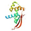
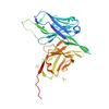
X-ray diffraction data for the Crystal structure of a large ribosomal subunit protein bL17 from Bordetella pertussis
First author:
Seattle Structural Genomics Center for Infectious Disease (SSGCID)
Uniprot: Q7VTA6
Resolution: 1.65 Å
R/Rfree: 0.19/0.22
Uniprot: Q7VTA6
Resolution: 1.65 Å
R/Rfree: 0.19/0.22
X-ray diffraction data for the psaea_00612_a_b3-36lw
First author:
Diffraction Data Upload SSGCID
X-ray diffraction data for the The dose series study of NcAA9D: wedge 20-38
First author:
Sam Miller
X-ray diffraction data for the The dose series study of NcAA9D: wedge 1–19
First author:
Sam Miller
X-ray diffraction data for the Crystal structure of hen egg white lysozyme at 100 Kelvin with Apiezon N

X-ray diffraction data for the Crystal structure of hen egg white lysozyme at 100 Kelvin with Apiezon N (Duplicate)
X-ray diffraction data for the Crystal structure of hen egg white lysozyme at 100 Kelvin with Lard (Triplicate)

X-ray diffraction data for the Crystal structure of hen egg white lysozyme at 100 Kelvin with Apiezon N (Triplicate)

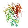
X-ray diffraction data for the Crystal structure of Phosphoribosylaminoimidazole carboxylase from Burkholderia xenovorans (Apo)
First author:
Seattle Structural Genomics Center for Infectious Disease (SSGCID)
Uniprot: Q13UJ9
Resolution: 2.39 Å
R/Rfree: 0.23/0.28
Uniprot: Q13UJ9
Resolution: 2.39 Å
R/Rfree: 0.23/0.28
X-ray diffraction data for the Crystal structure of the cysteine-free anti-UTag intrabody
First author:
S.H. Chen
Resolution: 1.45 Å
R/Rfree: 0.17/0.21
Resolution: 1.45 Å
R/Rfree: 0.17/0.21
X-ray diffraction data for the Crystal structure of human adenosine kinase (ADK) in complex with inhibitor BKI-1676
First author:
Seattle Structural Genomics Center for Infectious Disease (SSGCID)
Uniprot: P55263
Resolution: 2.81 Å
R/Rfree: 0.24/0.27
Uniprot: P55263
Resolution: 2.81 Å
R/Rfree: 0.24/0.27
X-ray diffraction data for the Crystal structure of human adenosine kinase (ADK) in complex with inhibitor BKI-1817
First author:
Seattle Structural Genomics Center for Infectious Disease (SSGCID)
Uniprot: P55263
Resolution: 2.51 Å
R/Rfree: 0.21/0.25
Uniprot: P55263
Resolution: 2.51 Å
R/Rfree: 0.21/0.25
X-ray diffraction data for the Crystal structure of human adenosine kinase (ADK) in complex with inhibitor BKI-1553

First author:
Seattle Structural Genomics Center for Infectious Disease (SSGCID)
Uniprot: P55263
Resolution: 2.41 Å
R/Rfree: 0.22/0.26
Uniprot: P55263
Resolution: 2.41 Å
R/Rfree: 0.22/0.26