6885 results
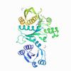
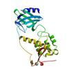
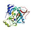
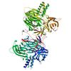
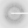
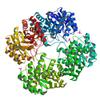
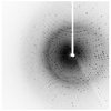
X-ray diffraction data for the Pseudomonas aeruginosa MurC with AZ5595
First author:
P.S. Horanyi
Resolution: 2.25 Å
R/Rfree: 0.18/0.22
Resolution: 2.25 Å
R/Rfree: 0.18/0.22
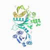
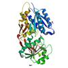
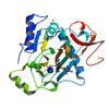
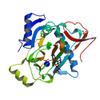
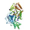
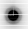
X-ray diffraction data for the Pseudomonas aeruginosa MurC with AZ8074
First author:
P.S. Horanyi
Resolution: 2.35 Å
R/Rfree: 0.18/0.22
Resolution: 2.35 Å
R/Rfree: 0.18/0.22
X-ray diffraction data for the Structure of mammalian NEIL2 from Monodelphis domestica
First author:
B.E. Eckenroth
Resolution: 2.54 Å
R/Rfree: 0.25/0.28
Resolution: 2.54 Å
R/Rfree: 0.25/0.28
X-ray diffraction data for the Crystal Structure of Leucine-, isoleucine-, valine-, threonine-, and alanine-binding protein from Brucella ovis, closed conformation
First author:
Seattle Structural Genomics Center for Infectious Disease (SSGCID)
Resolution: 1.70 Å
R/Rfree: 0.16/0.19
Resolution: 1.70 Å
R/Rfree: 0.16/0.19
X-ray diffraction data for the Crystal Structure of Argininosuccinate synthase from Legionella pneumophila Philadelphia 1
First author:
Seattle Structural Genomics Center for Infectious Disease (SSGCID)
Resolution: 1.95 Å
R/Rfree: 0.16/0.19
Resolution: 1.95 Å
R/Rfree: 0.16/0.19
X-ray diffraction data for the The crystal structure of kanamycin B dioxygenase (KanJ) from Streptomyces kanamyceticus complex with nickel, sulfate and chloride
First author:
B. Mrugala
Resolution: 2.50 Å
R/Rfree: 0.17/0.21
Resolution: 2.50 Å
R/Rfree: 0.17/0.21
X-ray diffraction data for the The crystal structure of kanamycin B dioxygenase (KanJ) from Streptomyces kanamyceticus in complex with nickel, ribostamycin B and 2-oxoglutarate
First author:
B. Mrugala
Resolution: 2.40 Å
R/Rfree: 0.17/0.20
Resolution: 2.40 Å
R/Rfree: 0.17/0.20
X-ray diffraction data for the The crystal structure of kanamycin B dioxygenase (KanJ) from Streptomyces kanamyceticus in complex with nickel, sulfate, soaked with iodide

First author:
B. Mrugala
Resolution: 2.10 Å
R/Rfree: 0.17/0.20
Resolution: 2.10 Å
R/Rfree: 0.17/0.20
X-ray diffraction data for the The crystal structure of kanamycin B dioxygenase (KanJ) from Streptomyces kanamyceticus in complex with nickel and 2-oxoglutarate
First author:
B. Mrugala
Resolution: 2.15 Å
R/Rfree: 0.20/0.24
Resolution: 2.15 Å
R/Rfree: 0.20/0.24
X-ray diffraction data for the The crystal structure of kanamycin B dioxygenase (KanJ) from Streptomyces kanamyceticus in complex with nickel, neamine and sulfate
First author:
B. Mrugala
Resolution: 3.00 Å
R/Rfree: 0.20/0.23
Resolution: 3.00 Å
R/Rfree: 0.20/0.23
X-ray diffraction data for the Crystal structure of the SUN1-KASH4 6:6 complex
First author:
M. Gurusaran
Resolution: 2.75 Å
R/Rfree: 0.22/0.26
Resolution: 2.75 Å
R/Rfree: 0.22/0.26
X-ray diffraction data for the The crystal structure of kanamycin B dioxygenase (KanJ) from Streptomyces kanamyceticus in complex with nickel and kanamycin B sulfate
First author:
B. Mrugala
Resolution: 2.36 Å
R/Rfree: 0.19/0.23
Resolution: 2.36 Å
R/Rfree: 0.19/0.23
X-ray diffraction data for the Crystal Structure of Dihydrodipicolinate synthase (DHDPS) from Brucella suis 1330
First author:
Seattle Structural Genomics Center for Infectious Disease (SSGCID)
Resolution: 2.20 Å
R/Rfree: 0.14/0.16
Resolution: 2.20 Å
R/Rfree: 0.14/0.16
X-ray diffraction data for the Crystal structure of Phosphoserine aminotransferase (SerC) from Stenotrophomonas maltophilia K279a
First author:
Seattle Structural Genomics Center for Infectious Disease (SSGCID)
Resolution: 1.60 Å
R/Rfree: 0.16/0.18
Resolution: 1.60 Å
R/Rfree: 0.16/0.18
X-ray diffraction data for the Crystal structure of the 2019-nCoV main protease complexed with GC376
First author:
L.F. Fu
Resolution: 1.40 Å
R/Rfree: 0.18/0.20
Resolution: 1.40 Å
R/Rfree: 0.18/0.20