6913 results
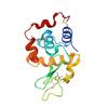
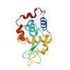
X-ray diffraction data for the Crystal structure of hen egg white lysozyme

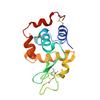
X-ray diffraction data for the Crystal structure of hen egg white lysozyme


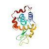
X-ray diffraction data for the Crystal structure of hen egg white lysozyme

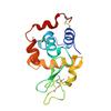
X-ray diffraction data for the Crystal structure of hen egg white lysozyme

X-ray diffraction data for the Crystal structure of hen egg white lysozyme


X-ray diffraction data for the Crystal structure of hen egg white lysozyme

X-ray diffraction data for the Crystal structure of hen egg white lysozyme


X-ray diffraction data for the Crystal structure of hen egg white lysozyme

X-ray diffraction data for the Crystal structure of hen egg white lysozyme


X-ray diffraction data for the Crystal structure of hen egg white lysozyme

