393 results
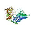
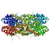
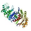
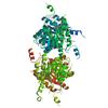
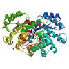
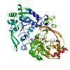
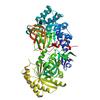
X-ray diffraction data for the 2.85 Angstrom resolution crystal structure of betaine aldehyde dehydrogenase (betB) H448F/P449M double mutant from Staphylococcus aureus in complex with NAD+ and BME-free Cys289
First author:
C. Chen
Gene name: betB
Resolution: 2.85 Å
R/Rfree: 0.16/0.21
Gene name: betB
Resolution: 2.85 Å
R/Rfree: 0.16/0.21
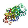
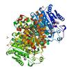
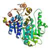
X-ray diffraction data for the Crystal Structure of K170M Mutant of Type I 3-Dehydroquinate Dehydratase (aroD) from Salmonella typhimurium LT2 in Non-Covalent Complex with Dehydroquinate.
First author:
S.H. Light
Gene name: aroD
Resolution: 1.60 Å
R/Rfree: 0.16/0.19
Gene name: aroD
Resolution: 1.60 Å
R/Rfree: 0.16/0.19
X-ray diffraction data for the Crystal structure of anabolic ornithine carbamoyltransferase from Vibrio vulnificus in complex with citrulline and inorganic phosphate
First author:
I.G. Shabalin
Resolution: 2.08 Å
R/Rfree: 0.16/0.19
Resolution: 2.08 Å
R/Rfree: 0.16/0.19
X-ray diffraction data for the Crystal structure of anabolic ornithine carbamoyltransferase from Vibrio vulnificus in complex with carbamoylphosphate and arginine
First author:
I.G. Shabalin
Resolution: 1.85 Å
R/Rfree: 0.15/0.19
Resolution: 1.85 Å
R/Rfree: 0.15/0.19
X-ray diffraction data for the Crystal structure of iron uptake ABC transporter substrate-binding protein PiaA from Streptococcus pneumoniae Canada MDR_19A bound to Bis-tris propane
First author:
P.J. Stogios
Resolution: 1.66 Å
R/Rfree: 0.17/0.19
Resolution: 1.66 Å
R/Rfree: 0.17/0.19
X-ray diffraction data for the Crystal structure of a putative Cas1 enzyme from Vibrio phage ICP1
First author:
A. Savchenko
Gene name: cas1
Resolution: 2.13 Å
R/Rfree: 0.17/0.21
Gene name: cas1
Resolution: 2.13 Å
R/Rfree: 0.17/0.21
X-ray diffraction data for the Crystal Structure of Type I 3-Dehydroquinate Dehydratase (aroD) from Salmonella typhimurium with close loop conformation.
First author:
S.H. Light
Gene name: aroD
Resolution: 1.90 Å
R/Rfree: 0.15/0.20
Gene name: aroD
Resolution: 1.90 Å
R/Rfree: 0.15/0.20
X-ray diffraction data for the 2.09 Angstrom resolution structure of a hypoxanthine-guanine phosphoribosyltransferase (hpt-1) from Bacillus anthracis str. 'Ames Ancestor' in complex with GMP
First author:
A.S. Halavaty
Gene name: hpt-1
Resolution: 2.09 Å
R/Rfree: 0.20/0.24
Gene name: hpt-1
Resolution: 2.09 Å
R/Rfree: 0.20/0.24
X-ray diffraction data for the 1.70 Angstrom resolution crystal structure of outer-membrane lipoprotein carrier protein (lolA) from Yersinia pestis CO92
First author:
A.S. Halavaty
Gene name: lolA
Resolution: 1.70 Å
R/Rfree: 0.20/0.24
Gene name: lolA
Resolution: 1.70 Å
R/Rfree: 0.20/0.24
X-ray diffraction data for the Crystal structure of spermidine N-acetyltransferase from Vibrio cholerae in complex with spermine
First author:
E.V. Filippova
Resolution: 1.85 Å
R/Rfree: 0.15/0.18
Resolution: 1.85 Å
R/Rfree: 0.15/0.18
X-ray diffraction data for the 1.95 Angstrom Resolution Crystal Structure of Epidermin Leader Peptide Processing Serine Protease (EpiP) S393A Mutant from Staphylococcus aureus
First author:
G. Minasov
Gene name: epiP
Resolution: 1.95 Å
R/Rfree: 0.17/0.20
Gene name: epiP
Resolution: 1.95 Å
R/Rfree: 0.17/0.20
X-ray diffraction data for the 2.4 Angstrom Crystal Structure of Dihydroorotase (pyrC) from Campylobacter jejuni.
First author:
G. Minasov
Gene name: pyrC
Resolution: 2.40 Å
R/Rfree: 0.19/0.25
Gene name: pyrC
Resolution: 2.40 Å
R/Rfree: 0.19/0.25
X-ray diffraction data for the 1.8 Angstrom resolution crystal structure of dihydroorotase (pyrC) from Salmonella enterica subsp. enterica serovar Typhimurium str. LT2
First author:
G. Minasov
Gene name: pyrC
Resolution: 1.80 Å
R/Rfree: 0.16/0.20
Gene name: pyrC
Resolution: 1.80 Å
R/Rfree: 0.16/0.20
X-ray diffraction data for the Crystal Structure of the 3-Dehydroquinate Dehydratase (aroD) from Salmonella typhimurium LT2 with Citrate Bound to the Active Site.
First author:
G. Minasov
Gene name: aroD
Resolution: 1.65 Å
R/Rfree: 0.16/0.20
Gene name: aroD
Resolution: 1.65 Å
R/Rfree: 0.16/0.20
X-ray diffraction data for the Crystal structure of unliganded anabolic ornithine carbamoyltransferase from Vibrio vulnificus at 1.86 A resolution
First author:
I.G. Shabalin
Resolution: 1.86 Å
R/Rfree: 0.16/0.20
Resolution: 1.86 Å
R/Rfree: 0.16/0.20
X-ray diffraction data for the Crystal Structure of a glutamate-1-semialdehyde aminotransferase from Bacillus anthracis with bound Pyridoxal 5'Phosphate
First author:
S.S. Sharma
Gene name: hemL-2
Resolution: 1.95 Å
R/Rfree: 0.15/0.19
Gene name: hemL-2
Resolution: 1.95 Å
R/Rfree: 0.15/0.19
X-ray diffraction data for the Crystal structure of the aminoglycoside phosphotransferase APH(3')-Ia, with substrate kanamycin and small molecule inhibitor tyrphostin AG1478

First author:
P.J. Stogios
Gene name: aphA1
Resolution: 2.71 Å
R/Rfree: 0.18/0.24
Gene name: aphA1
Resolution: 2.71 Å
R/Rfree: 0.18/0.24
X-ray diffraction data for the 1.55 Angstrom Crystal Structure of the Four Helical Bundle Membrane Localization Domain (4HBM) of the Vibrio vulnificus MARTX Effector Domain DUF5
First author:
G. Minasov
Resolution: 1.55 Å
R/Rfree: 0.17/0.22
Resolution: 1.55 Å
R/Rfree: 0.17/0.22
X-ray diffraction data for the Crystal structure of beta-ketoacyl-ACP synthase III (FabH) from Vibrio Cholerae in complex with Coenzyme A

First author:
J. Hou
Resolution: 1.78 Å
R/Rfree: 0.17/0.20
Resolution: 1.78 Å
R/Rfree: 0.17/0.20
X-ray diffraction data for the 1.4A Crystal Structure of Isocitrate Lyase from Yersinia pestis CO92
First author:
S.S. Sharma
Gene name: aceA
Resolution: 1.40 Å
R/Rfree: 0.13/0.17
Gene name: aceA
Resolution: 1.40 Å
R/Rfree: 0.13/0.17
X-ray diffraction data for the 1.4 Angstrom Resolution Crystal Structure of Putative alpha Amylase from Salmonella typhimurium.
First author:
G. Minasov
Resolution: 1.40 Å
R/Rfree: 0.15/0.17
Resolution: 1.40 Å
R/Rfree: 0.15/0.17
X-ray diffraction data for the 1.8 Angstrom Resolution Crystal Structure of Enoyl-CoA Hydratase from Bacillus anthracis.
First author:
G. Minasov
Resolution: 1.80 Å
R/Rfree: 0.16/0.19
Resolution: 1.80 Å
R/Rfree: 0.16/0.19
X-ray diffraction data for the Crystal Structure of a Putative Macrophage Growth Locus, subunit A From Francisella tularensis SCHU S4

First author:
J.S. Brunzelle
Gene name: mglA
Resolution: 2.75 Å
R/Rfree: 0.19/0.23
Gene name: mglA
Resolution: 2.75 Å
R/Rfree: 0.19/0.23
X-ray diffraction data for the 1.95 Angstrom crystal structure of a bifunctional 3-deoxy-7-phosphoheptulonate synthase/chorismate mutase (aroA) from Listeria monocytogenes EGD-e
First author:
S.H. Light
Gene name: aroA
Resolution: 1.95 Å
R/Rfree: 0.15/0.20
Gene name: aroA
Resolution: 1.95 Å
R/Rfree: 0.15/0.20
X-ray diffraction data for the Crystal Structure of Short Chain Dehydrogenase (yciK) from Salmonella enterica subsp. enterica serovar Typhimurium str. LT2 in Complex with NADP and Acetate.
First author:
G. Minasov
Gene name: yciK
Resolution: 1.83 Å
R/Rfree: 0.14/0.18
Gene name: yciK
Resolution: 1.83 Å
R/Rfree: 0.14/0.18
X-ray diffraction data for the 2.7 Angstrom resolution crystal structure of a probable holliday junction DNA helicase (ruvB) from Campylobacter jejuni subsp. jejuni NCTC 11168 in complex with adenosine-5'-diphosphate

First author:
A.S. Halavaty
Gene name: ruvB
Resolution: 2.69 Å
R/Rfree: 0.22/0.27
Gene name: ruvB
Resolution: 2.69 Å
R/Rfree: 0.22/0.27
X-ray diffraction data for the Crystal structure of the aminoglycoside phosphotransferase APH(3')-Ia, with substrate kanamycin and small molecule inhibitor pyrazolopyrimidine PP1
First author:
P.J. Stogios
Gene name: aphA1
Resolution: 1.89 Å
R/Rfree: 0.15/0.20
Gene name: aphA1
Resolution: 1.89 Å
R/Rfree: 0.15/0.20
X-ray diffraction data for the 2.1 Angstrom resolution crystal structure of uncharacterized protein lmo0859 from Listeria monocytogenes EGD-e
First author:
A.S. Halavaty
Gene name: -
Resolution: 2.10 Å
R/Rfree: 0.17/0.21
Gene name: -
Resolution: 2.10 Å
R/Rfree: 0.17/0.21
X-ray diffraction data for the 2.06 Angstrom resolution structure of a hypoxanthine-guanine phosphoribosyltransferase (hpt-1) from Bacillus anthracis str. 'Ames Ancestor'
First author:
A.S. Halavaty
Gene name: hpt-1
Resolution: 2.06 Å
R/Rfree: 0.17/0.21
Gene name: hpt-1
Resolution: 2.06 Å
R/Rfree: 0.17/0.21
X-ray diffraction data for the 2.65 Angstrom resolution crystal structure of an orotate phosphoribosyltransferase from Bacillus anthracis str. 'Ames Ancestor' in complex with 5-phospho-alpha-D-ribosyl diphosphate (PRPP)
First author:
A.S. Halavaty
Gene name: pyrE
Resolution: 2.65 Å
R/Rfree: 0.22/0.26
Gene name: pyrE
Resolution: 2.65 Å
R/Rfree: 0.22/0.26
X-ray diffraction data for the Structural flexibility in region involved in dimer formation of nuclease domain of Ribonuclase III (rnc) from Campylobacter jejuni
First author:
G. Minasov
Gene name: rnc
Resolution: 1.25 Å
R/Rfree: 0.13/0.17
Gene name: rnc
Resolution: 1.25 Å
R/Rfree: 0.13/0.17
X-ray diffraction data for the Crystal structure of a putative succinate-semialdehyde dehydrogenase from salmonella typhimurium lt2
First author:
H. Zheng
Gene name: yneI
Resolution: 1.85 Å
R/Rfree: 0.17/0.19
Gene name: yneI
Resolution: 1.85 Å
R/Rfree: 0.17/0.19
X-ray diffraction data for the Crystal structure of BA2930 mutant (H183A) in complex with AcCoA
First author:
M.M. Klimecka
Gene name: aacC7
Resolution: 2.15 Å
R/Rfree: 0.17/0.23
Gene name: aacC7
Resolution: 2.15 Å
R/Rfree: 0.17/0.23
X-ray diffraction data for the 1.77 Angstrom resolution crystal structure of orotidine 5'-phosphate decarboxylase from Vibrio cholerae O1 biovar eltor str. N16961
First author:
A.S. Halavaty
Resolution: 1.77 Å
R/Rfree: 0.15/0.19
Resolution: 1.77 Å
R/Rfree: 0.15/0.19
X-ray diffraction data for the 2.8 Angstrom Crystal Structure of Sensor Domain of Histidine Kinase from Clostridium perfringens.
First author:
G. Minasov
Resolution: 2.80 Å
R/Rfree: 0.20/0.25
Resolution: 2.80 Å
R/Rfree: 0.20/0.25
X-ray diffraction data for the Crystal structure of the aminoglycoside phosphotransferase APH(3')-Ia, with substrate kanamycin and small molecule inhibitor 1-NA-PP1
First author:
P.J. Stogios
Gene name: aphA1
Resolution: 1.86 Å
R/Rfree: 0.16/0.22
Gene name: aphA1
Resolution: 1.86 Å
R/Rfree: 0.16/0.22
X-ray diffraction data for the Crystal structure of an amino acid ABC transporter substrate-binding protein from Streptococcus pneumoniae Canada MDR_19A bound to L-arginine, form 2
First author:
P.J. Stogios
Resolution: 1.78 Å
R/Rfree: 0.16/0.19
Resolution: 1.78 Å
R/Rfree: 0.16/0.19
X-ray diffraction data for the 1.90 Angstrom resolution crystal structure of betaine aldehyde dehydrogenase (betB) from Staphylococcus aureus in complex with NAD+ and BME-free Cys289
First author:
A.S. Halavaty
Gene name: betB
Resolution: 1.90 Å
R/Rfree: 0.14/0.19
Gene name: betB
Resolution: 1.90 Å
R/Rfree: 0.14/0.19
X-ray diffraction data for the Crystal structure of anabolic ornithine carbamoyltransferase from Vibrio vulnificus in complex with carbamoyl phosphate and L-norvaline
First author:
I.G. Shabalin
Resolution: 1.70 Å
R/Rfree: 0.14/0.17
Resolution: 1.70 Å
R/Rfree: 0.14/0.17
X-ray diffraction data for the 2.35 Angstrom resolution crystal structure of putative O-acetylhomoserine (thiol)-lyase (metY) from Campylobacter jejuni subsp. jejuni NCTC 11168 with N'-Pyridoxyl-Lysine-5'-Monophosphate at position 205

First author:
A.S. Halavaty
Gene name: metY
Resolution: 2.35 Å
R/Rfree: 0.20/0.25
Gene name: metY
Resolution: 2.35 Å
R/Rfree: 0.20/0.25
X-ray diffraction data for the 2.1 Angstrom resolution crystal structure of glycerol-3-phosphate dehydrogenase (gpsA) from Coxiella burnetii
First author:
G. Minasov
Gene name: gpsA
Resolution: 2.10 Å
R/Rfree: 0.16/0.21
Gene name: gpsA
Resolution: 2.10 Å
R/Rfree: 0.16/0.21
X-ray diffraction data for the Crystal Structure of Leukotoxin (LukE) from Staphylococcus aureus subsp. aureus COL.
First author:
S. Nocadello
Gene name: lukE
Resolution: 3.20 Å
R/Rfree: 0.18/0.22
Gene name: lukE
Resolution: 3.20 Å
R/Rfree: 0.18/0.22
X-ray diffraction data for the 1.42 Angstrom resolution crystal structure of accessory colonization factor AcfC (acfC) in complex with D-aspartic acid
First author:
A.S. Halavaty
Gene name: acfC
Resolution: 1.42 Å
R/Rfree: 0.13/0.14
Gene name: acfC
Resolution: 1.42 Å
R/Rfree: 0.13/0.14
X-ray diffraction data for the Crystal structure of the aminoglycoside phosphotransferase APH(3')-Ia, with substrate kanamycin and small molecule inhibitor anthrapyrazolone SP600125
First author:
P.J. Stogios
Gene name: aphA1
Resolution: 2.37 Å
R/Rfree: 0.16/0.22
Gene name: aphA1
Resolution: 2.37 Å
R/Rfree: 0.16/0.22
X-ray diffraction data for the 2.2 Angstrom Crystal Structure of Conserved Hypothetical Protein from Bacillus anthracis.
First author:
G. Minasov
Resolution: 2.20 Å
R/Rfree: 0.18/0.21
Resolution: 2.20 Å
R/Rfree: 0.18/0.21
X-ray diffraction data for the Coproporphyrinogen III oxidase (HemF) from Acinetobacter baumannii
First author:
J. Abendroth
Resolution: 1.45 Å
R/Rfree: 0.14/0.17
Resolution: 1.45 Å
R/Rfree: 0.14/0.17
X-ray diffraction data for the Crystal structure of glutathione S-transferase from Sinorhizobium meliloti 1021, NYSGRC target 021389
First author:
I.G. Shabalin
Resolution: 1.78 Å
R/Rfree: 0.16/0.19
Resolution: 1.78 Å
R/Rfree: 0.16/0.19
X-ray diffraction data for the 1.52 Angstrom Crystal Structure of A42R Profilin-like Protein from Monkeypox Virus Zaire-96-I-16
First author:
G. Minasov
Gene name: A42R
Resolution: 1.52 Å
R/Rfree: 0.14/0.17
Gene name: A42R
Resolution: 1.52 Å
R/Rfree: 0.14/0.17
X-ray diffraction data for the Crystal structure of a type VI secretion system effector from Yersinia pestis
First author:
E.V. Filippova
Resolution: 2.10 Å
R/Rfree: 0.22/0.27
Resolution: 2.10 Å
R/Rfree: 0.22/0.27
X-ray diffraction data for the 1.85 Angstrom Resolution Crystal Structure of PTS System Cellobiose-specific Transporter Subunit IIB from Bacillus anthracis.
First author:
G. Minasov
Gene name: celA-2
Resolution: 1.85 Å
R/Rfree: 0.18/0.22
Gene name: celA-2
Resolution: 1.85 Å
R/Rfree: 0.18/0.22
X-ray diffraction data for the 1.95 Angstrom Crystal Structure of Gamma-glutamyl phosphate Reductase from Saccharomonospora viridis.
First author:
G. Minasov
Resolution: 1.95 Å
R/Rfree: 0.17/0.21
Resolution: 1.95 Å
R/Rfree: 0.17/0.21
X-ray diffraction data for the 3.0 Angstrom resolution crystal structure of dTDP-glucose 4,6-dehydratase (rfbB) from Bacillus anthracis str. Ames in complex with NAD

First author:
A.S. Halavaty
Gene name: rfbB
Resolution: 3.00 Å
R/Rfree: 0.17/0.23
Gene name: rfbB
Resolution: 3.00 Å
R/Rfree: 0.17/0.23
X-ray diffraction data for the 2.27 Angstrom Crystal Structure of beta-Phosphoglucomutase (pgmB) from Clostridium difficile
First author:
G. Minasov
Gene name: pgmB
Resolution: 2.27 Å
R/Rfree: 0.19/0.26
Gene name: pgmB
Resolution: 2.27 Å
R/Rfree: 0.19/0.26
X-ray diffraction data for the 1.95 Angstrom Resolution Crystal Structure of 3-deoxy-D-manno-octulosonate 8-phosphate phosphatase from Yersinia pestis
First author:
G. Minasov
Resolution: 1.95 Å
R/Rfree: 0.16/0.20
Resolution: 1.95 Å
R/Rfree: 0.16/0.20
X-ray diffraction data for the Crystal structure of BaLdcB / VanY-like L,D-carboxypeptidase Zinc(II)-free

First author:
C.N. Hoyland
Resolution: 2.30 Å
R/Rfree: 0.19/0.26
Resolution: 2.30 Å
R/Rfree: 0.19/0.26
X-ray diffraction data for the 2.05 Angstrom Crystal Structure of Putative 5'-Nucleotidase from Staphylococcus aureus in complex with alpha-ketoglutarate
First author:
G. Minasov
Resolution: 2.05 Å
R/Rfree: 0.17/0.22
Resolution: 2.05 Å
R/Rfree: 0.17/0.22
X-ray diffraction data for the 2.0 Angstrom Resolution Crystal Structure of Glucose-6-phosphate Isomerase (pgi) from Bacillus anthracis.
First author:
G. Minasov
Gene name: pgi
Resolution: 2.00 Å
R/Rfree: 0.14/0.19
Gene name: pgi
Resolution: 2.00 Å
R/Rfree: 0.14/0.19
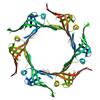
X-ray diffraction data for the Phosphoribosylaminoimidazole carboxylase with fructose-6-phosphate bound to the central channel of the octameric protein structure.
First author:
E.V. Filippova
Gene name: purE
Resolution: 2.00 Å
R/Rfree: 0.19/0.24
Gene name: purE
Resolution: 2.00 Å
R/Rfree: 0.19/0.24
X-ray diffraction data for the 2.4 Angstrom resolution crystal structure of shikimate 5-dehydrogenase (aroE) from Vibrio cholerae O1 biovar eltor str. N16961 in complex with shikimate and NADPH
First author:
A.S. Halavaty
Gene name: aroE
Resolution: 2.40 Å
R/Rfree: 0.24/0.28
Gene name: aroE
Resolution: 2.40 Å
R/Rfree: 0.24/0.28
X-ray diffraction data for the Structure of a putative transcriptional regulator of LacI family from Sanguibacter keddieii DSM 10542.
First author:
E.V. Filippova
Resolution: 1.45 Å
R/Rfree: 0.13/0.18
Resolution: 1.45 Å
R/Rfree: 0.13/0.18
X-ray diffraction data for the Biosynthetic Thiolase (ThlA1) from Clostridium difficile
First author:
E.V. Filippova
Gene name: thlA1
Resolution: 1.25 Å
R/Rfree: 0.12/0.14
Gene name: thlA1
Resolution: 1.25 Å
R/Rfree: 0.12/0.14
X-ray diffraction data for the 2.35 Angstrom resolution crystal structure of a putative tRNA (guanine-7-)-methyltransferase (trmD) from Staphylococcus aureus subsp. aureus MRSA252

First author:
A.S. Halavaty
Gene name: trmD
Resolution: 2.35 Å
R/Rfree: 0.21/0.26
Gene name: trmD
Resolution: 2.35 Å
R/Rfree: 0.21/0.26
X-ray diffraction data for the 2.6 Angstrom Crystal Structure of Putative yceG-like Protein lmo1499 from Listeria monocytogenes
First author:
G. Minasov
Gene name: -
Resolution: 2.60 Å
R/Rfree: 0.17/0.22
Gene name: -
Resolution: 2.60 Å
R/Rfree: 0.17/0.22
X-ray diffraction data for the Crystal structure of peptidoglycan glycosyltransferase from Atopobium parvulum DSM 20469.
First author:
E.V. Filippova
Resolution: 1.92 Å
R/Rfree: 0.20/0.24
Resolution: 1.92 Å
R/Rfree: 0.20/0.24
X-ray diffraction data for the Crystal structure of type III effector protein ExoU (exoU)
First author:
G.H. Tyson
Gene name: exoU
Resolution: 2.50 Å
R/Rfree: 0.21/0.27
Gene name: exoU
Resolution: 2.50 Å
R/Rfree: 0.21/0.27
X-ray diffraction data for the X-ray Crystal Structure of a Putative Lipoprotein from Bacillus anthracis
First author:
J.S. Brunzelle
Resolution: 2.42 Å
R/Rfree: 0.18/0.21
Resolution: 2.42 Å
R/Rfree: 0.18/0.21
X-ray diffraction data for the Crystal structure of hypothetical protein with ketosteroid isomerase-like protein fold from Catenulispora acidiphila DSM 44928
First author:
E.V. Filippova
Resolution: 1.15 Å
R/Rfree: 0.13/0.14
Resolution: 1.15 Å
R/Rfree: 0.13/0.14
X-ray diffraction data for the 1.75 Angstrom Crystal Structure of Transcriptional Regulator ftom Vibrio vulnificus.
First author:
G. Minasov
Resolution: 1.75 Å
R/Rfree: 0.18/0.22
Resolution: 1.75 Å
R/Rfree: 0.18/0.22
X-ray diffraction data for the Crystal structure of glyoxalase/bleomycin resistance protein from Catenulispora acidiphila.

First author:
E.V. Filippova
Resolution: 1.73 Å
R/Rfree: 0.17/0.19
Resolution: 1.73 Å
R/Rfree: 0.17/0.19
X-ray diffraction data for the Structure of phage-related protein from Bacillus cereus ATCC 10987
First author:
E.V. Filippova
Resolution: 1.30 Å
R/Rfree: 0.12/0.16
Resolution: 1.30 Å
R/Rfree: 0.12/0.16
X-ray diffraction data for the 2.06 Angstrom resolution crystal structure of phosphomethylpyrimidine kinase (thiD)from Clostridium difficile 630
First author:
A.S. Halavaty
Gene name: thiD
Resolution: 2.06 Å
R/Rfree: 0.18/0.22
Gene name: thiD
Resolution: 2.06 Å
R/Rfree: 0.18/0.22
X-ray diffraction data for the Crystal structure of hypothetical protein with ketosteroid isomerase-like protein fold from Catenulispora acidiphila DSM 44928 in complex with Trimethylamine.
First author:
E.V. Filippova
Resolution: 1.95 Å
R/Rfree: 0.18/0.22
Resolution: 1.95 Å
R/Rfree: 0.18/0.22
X-ray diffraction data for the 1.45 Angstrom Crystal Structure of Shikimate 5-dehydrogenase from Listeria monocytogenes in Complex with Shikimate and NAD.
First author:
G. Minasov
Gene name: -
Resolution: 1.45 Å
R/Rfree: 0.15/0.18
Gene name: -
Resolution: 1.45 Å
R/Rfree: 0.15/0.18
X-ray diffraction data for the 1.03 Angstrom Crystal Structure of Q236A Mutant Type I Dehydroquinate Dehydratase (aroD) from Salmonella typhimurium
First author:
S.H. Light
Gene name: aroD
Resolution: 1.03 Å
R/Rfree: 0.14/0.16
Gene name: aroD
Resolution: 1.03 Å
R/Rfree: 0.14/0.16
X-ray diffraction data for the Beta-ketoacyl-acyl carrier protein synthase III-2 (FabH2)(C113A) from Vibrio cholerae
First author:
J. Hou
Resolution: 1.61 Å
R/Rfree: 0.15/0.18
Resolution: 1.61 Å
R/Rfree: 0.15/0.18
X-ray diffraction data for the Crystal structure of 3-ketoacyl-(acyl-carrier-protein) reductase (FabG) (G141A) from Vibrio cholerae in complex with NADPH
First author:
J. Hou
Gene name: fabG
Resolution: 2.21 Å
R/Rfree: 0.21/0.24
Gene name: fabG
Resolution: 2.21 Å
R/Rfree: 0.21/0.24
X-ray diffraction data for the Crystal structure of Thymidylate Kinase from Staphylococcus aureus in complex with 3'-Azido-3'-Deoxythymidine-5'-Monophosphate
First author:
E.V. Filippova
Resolution: 1.85 Å
R/Rfree: 0.18/0.23
Resolution: 1.85 Å
R/Rfree: 0.18/0.23
X-ray diffraction data for the Crystal structure of an aminoglycoside acetyltransferase HMB0005 from an uncultured soil metagenomic sample, unknown active site density modeled as polyethylene glycol
First author:
Z. Xu
Gene name: None
Resolution: 2.02 Å
R/Rfree: 0.19/0.22
Gene name: None
Resolution: 2.02 Å
R/Rfree: 0.19/0.22
X-ray diffraction data for the Crystal structure of Escherichia coli protein YodA in complex with Ni - artifact of purification.
First author:
O.A. Gasiorowska
Resolution: 1.95 Å
R/Rfree: 0.17/0.22
Resolution: 1.95 Å
R/Rfree: 0.17/0.22
X-ray diffraction data for the 2.1 Angstrom Resolution Crystal Structure of Metallo-beta-lactamase from Staphylococcus aureus subsp. aureus COL
First author:
G. Minasov
Resolution: 2.10 Å
R/Rfree: 0.16/0.20
Resolution: 2.10 Å
R/Rfree: 0.16/0.20
X-ray diffraction data for the Crystal structure of beta-ketoacyl-acyl carrier protein reductase (FabG)(G141A) from Vibrio cholerae
First author:
J. Hou
Gene name: fabG
Resolution: 2.40 Å
R/Rfree: 0.20/0.24
Gene name: fabG
Resolution: 2.40 Å
R/Rfree: 0.20/0.24
X-ray diffraction data for the 1.55 Angstrom Resolution Crystal Structure of Peptidase T (pepT-1) from Bacillus anthracis str. 'Ames Ancestor'.
First author:
G. Minasov
Gene name: pepT-1
Resolution: 1.55 Å
R/Rfree: 0.15/0.17
Gene name: pepT-1
Resolution: 1.55 Å
R/Rfree: 0.15/0.17
X-ray diffraction data for the 1.95 Angstrom Crystal Structure of of Type I 3-Dehydroquinate Dehydratase (aroD) from Clostridium difficile with Covalent Modified Comenic Acid.
First author:
G. Minasov
Gene name: aroD
Resolution: 1.95 Å
R/Rfree: 0.16/0.20
Gene name: aroD
Resolution: 1.95 Å
R/Rfree: 0.16/0.20
X-ray diffraction data for the 2.05 Angstrom resolution crystal structure of a short chain dehydrogenase from Bacillus anthracis str. 'Ames Ancestor' in complex with NAD+
First author:
A.S. Halavaty
Resolution: 2.05 Å
R/Rfree: 0.16/0.19
Resolution: 2.05 Å
R/Rfree: 0.16/0.19
X-ray diffraction data for the Crystal Structure of a pyridoxamine kinase from Yersinia pestis CO92
First author:
J.S. Brunzelle
Gene name: pdxY
Resolution: 1.89 Å
R/Rfree: 0.17/0.20
Gene name: pdxY
Resolution: 1.89 Å
R/Rfree: 0.17/0.20
X-ray diffraction data for the 1.8 Angstrom Crystal Structure of the N-terminal Domain of Protein with Unknown Function from Vibrio cholerae.
First author:
G. Minasov
Resolution: 1.80 Å
R/Rfree: 0.16/0.20
Resolution: 1.80 Å
R/Rfree: 0.16/0.20
X-ray diffraction data for the 2.17 Angstrom resolution crystal structure of malate dehydrogenase from Vibrio vulnificus CMCP6

First author:
A.S. Halavaty
Resolution: 2.17 Å
R/Rfree: 0.16/0.21
Resolution: 2.17 Å
R/Rfree: 0.16/0.21
X-ray diffraction data for the Crystal structure of phosphate ABC transporter, periplasmic phosphate-binding protein PstS 1 (PBP1) from Streptococcus pneumoniae Canada MDR_19A
First author:
P.J. Stogios
Resolution: 1.70 Å
R/Rfree: 0.17/0.21
Resolution: 1.70 Å
R/Rfree: 0.17/0.21
X-ray diffraction data for the 1.65 Angstrom resolution crystal structure of betaine aldehyde dehydrogenase (betB) from Staphylococcus aureus with BME-modified Cys289 and PEG molecule in active site
First author:
A.S. Halavaty
Gene name: betB
Resolution: 1.65 Å
R/Rfree: 0.14/0.16
Gene name: betB
Resolution: 1.65 Å
R/Rfree: 0.14/0.16
X-ray diffraction data for the 2.06 Angstrom resolution crystal structure of a short chain dehydrogenase from Bacillus anthracis str. 'Ames Ancestor' in complex with NAD-acetone
First author:
A.S. Halavaty
Resolution: 2.06 Å
R/Rfree: 0.16/0.21
Resolution: 2.06 Å
R/Rfree: 0.16/0.21
X-ray diffraction data for the 2.25 Angstrom Crystal Structure of Phosphoserine Aminotransferase (SerC) from Salmonella enterica subsp. enterica serovar Typhimurium
First author:
G. Minasov
Gene name: serC
Resolution: 2.25 Å
R/Rfree: 0.19/0.24
Gene name: serC
Resolution: 2.25 Å
R/Rfree: 0.19/0.24
X-ray diffraction data for the 1.0 Angstrom resolution crystal structure of the branched-chain amino acid transporter substrate binding protein LivJ from Streptococcus pneumoniae str. Canada MDR_19A in complex with Isoleucine
First author:
A.S. Halavaty
Resolution: 1.00 Å
R/Rfree: 0.13/0.15
Resolution: 1.00 Å
R/Rfree: 0.13/0.15
X-ray diffraction data for the 2.25 Angstrom resolution crystal structure of a thymidylate kinase (tmk) from Vibrio cholerae O1 biovar eltor str. N16961 in complex with thymidine
First author:
A.S. Halavaty
Gene name: tmk
Resolution: 2.25 Å
R/Rfree: 0.21/0.26
Gene name: tmk
Resolution: 2.25 Å
R/Rfree: 0.21/0.26
X-ray diffraction data for the Crystal structure of BA2930 in complex with AcCoA and cytosine

First author:
M.M. Klimecka
Gene name: aacC7
Resolution: 2.00 Å
R/Rfree: 0.17/0.23
Gene name: aacC7
Resolution: 2.00 Å
R/Rfree: 0.17/0.23
X-ray diffraction data for the 1.99 Angstrom resolution crystal structure of a short chain dehydrogenase from Bacillus anthracis str. 'Ames Ancestor'
First author:
A.S. Halavaty
Resolution: 1.99 Å
R/Rfree: 0.17/0.21
Resolution: 1.99 Å
R/Rfree: 0.17/0.21
X-ray diffraction data for the 1.8 Angstrom Resolution Crystal Structure of Diaminopimelate Decarboxylase (lysA) from Vibrio cholerae.
First author:
G. Minasov
Resolution: 1.80 Å
R/Rfree: 0.18/0.22
Resolution: 1.80 Å
R/Rfree: 0.18/0.22
X-ray diffraction data for the 2.25 Angstrom resolution crystal structure of UDP-N-acetylmuramate--L-alanine ligase (murC) from Yersinia pestis CO92 in complex with AMP
First author:
A.S. Halavaty
Gene name: murC
Resolution: 2.25 Å
R/Rfree: 0.18/0.22
Gene name: murC
Resolution: 2.25 Å
R/Rfree: 0.18/0.22
X-ray diffraction data for the 1.95 Angstrom Crystal Structure of Complex of Hypoxanthine-Guanine Phosphoribosyltransferase from Bacillus anthracis with 2-(N-morpholino)ethanesulfonic acid (MES)
First author:
G. Minasov
Gene name: hpt-1
Resolution: 1.95 Å
R/Rfree: 0.19/0.23
Gene name: hpt-1
Resolution: 1.95 Å
R/Rfree: 0.19/0.23
X-ray diffraction data for the Crystal structure of iron uptake ABC transporter substrate-binding protein PiuA from Streptococcus pneumoniae Canada MDR_19A

First author:
P.J. Stogios
Resolution: 1.13 Å
R/Rfree: 0.15/0.17
Resolution: 1.13 Å
R/Rfree: 0.15/0.17
X-ray diffraction data for the Alpha-Helical barrel formed by the decamer of the zinc resistance-associated protein (STM4172) from Salmonella enterica subsp. enterica serovar Typhimurium str. LT2
First author:
E.V. Filippova
Gene name: zraP
Resolution: 2.70 Å
R/Rfree: 0.22/0.27
Gene name: zraP
Resolution: 2.70 Å
R/Rfree: 0.22/0.27