1160 results
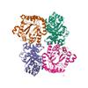
X-ray diffraction data for the Crystal structure of the catalytic domain of DapE protein from V.cholerea in the Zn bound form
First author:
M.Makowska-Grzyska 'B.Nocek
Resolution: 1.65 Å
R/Rfree: 0.16/0.18
Resolution: 1.65 Å
R/Rfree: 0.16/0.18
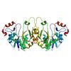
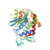
X-ray diffraction data for the 1.85 Angstrom Crystal Structure of GCPE Protein from Bacillus anthracis
First author:
G. Minasov
Resolution: 1.85 Å
R/Rfree: 0.13/0.17
Resolution: 1.85 Å
R/Rfree: 0.13/0.17
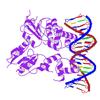
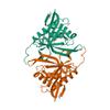
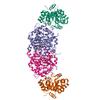
X-ray diffraction data for the Structure of yncA, a putative ACETYLTRANSFERASE from Salmonella typhimurium
First author:
A.U. Singer
Gene name: argC
Resolution: 1.75 Å
R/Rfree: 0.16/0.20
Gene name: argC
Resolution: 1.75 Å
R/Rfree: 0.16/0.20
X-ray diffraction data for the Crystal structure of the catalytic domain of DapE protein from V.cholerea in the Zn bound form
First author:
B. Nocek
Resolution: 1.65 Å
R/Rfree: 0.14/0.17
Resolution: 1.65 Å
R/Rfree: 0.14/0.17
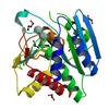
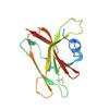
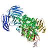
X-ray diffraction data for the 1.4 Angstrom resolution crystal structure of uncharacterized protein BA_2500 from Bacillus anthracis str. Ames
First author:
A.S. Halavaty
Resolution: 1.40 Å
R/Rfree: 0.15/0.19
Resolution: 1.40 Å
R/Rfree: 0.15/0.19
X-ray diffraction data for the Crystal structure of tryptophan synthase from M. tuberculosis - ligand-free form
X-ray diffraction data for the Cycloalternan-forming enzyme from Listeria monocytogenes in complex with cycloalternan
X-ray diffraction data for the 2.95 Angstrom Crystal Structure of the Dimeric Form of Penicillin Binding Protein 2 Prime from Enterococcus faecium

X-ray diffraction data for the 1.9 Angstrom Crystal Structure of 3-deoxy-manno-octulosonate Cytidylyltransferase (kdsB) from Acinetobacter baumannii without His-Tag Bound to the Active Site