1160 results
X-ray diffraction data for the Crystal Structure of Protein of Unknown Function YPO2564 from Yersinia pestis
First author:
Y. Kim
Gene name: yicN
Resolution: 2.00 Å
R/Rfree: 0.20/0.22
Gene name: yicN
Resolution: 2.00 Å
R/Rfree: 0.20/0.22
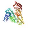
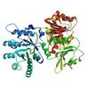
X-ray diffraction data for the Crystal structure of YPT31, a Rab family GTPase from Candida albicans, in complex with GDP and Zn(II)

First author:
P.J. Stogios
Gene name: None
Resolution: 2.35 Å
R/Rfree: 0.19/0.23
Gene name: None
Resolution: 2.35 Å
R/Rfree: 0.19/0.23
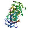
X-ray diffraction data for the Crystal structure of the phosphomannomutase PMM1 from Candida albicans, apoenzyme state
First author:
P.J. Stogios
Gene name: None
Resolution: 1.95 Å
R/Rfree: 0.21/0.26
Gene name: None
Resolution: 1.95 Å
R/Rfree: 0.21/0.26
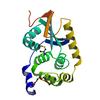
X-ray diffraction data for the Crystal structure of tryptophan synthase from M. tuberculosis - aminoacrylate-bound form
X-ray diffraction data for the Crystal structure of Equine Serum Albumin complex with etodolac
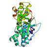
X-ray diffraction data for the Crystal structure of adenylosuccinate lyase ADE13 from Candida albicans
First author:
P.J. Stogios
Gene name: None
Resolution: 2.00 Å
R/Rfree: 0.15/0.19
Gene name: None
Resolution: 2.00 Å
R/Rfree: 0.15/0.19
X-ray diffraction data for the MdbA protein, a thiol-disulfide oxidoreductase from Corynebacterium matruchotii
First author:
J. Osipiuk
Resolution: 1.20 Å
R/Rfree: 0.12/0.15
Resolution: 1.20 Å
R/Rfree: 0.12/0.15
X-ray diffraction data for the Dehydroquinate dehydratase and shikimate dehydrogenase from S. pombe AroM
X-ray diffraction data for the Putative Antitoxin HicB3 from Escherichia coli str. K-12 substr. DH10B
First author:
J. Osipiuk
Gene name: ydcQ
Resolution: 1.90 Å
R/Rfree: 0.20/0.25
Gene name: ydcQ
Resolution: 1.90 Å
R/Rfree: 0.20/0.25
X-ray diffraction data for the OmpA-like domain of FopA1 from Francisella tularensis subsp. tularensis SCHU S4
First author:
K. Michalska
Gene name: fopA1
Resolution: 2.36 Å
R/Rfree: 0.21/0.24
Gene name: fopA1
Resolution: 2.36 Å
R/Rfree: 0.21/0.24
X-ray diffraction data for the Crystal Structure of NSP15 Endoribonuclease from SARS CoV-2.
First author:
Y. Kim
Gene name: orf1ab
Resolution: 2.20 Å
R/Rfree: 0.16/0.18
Gene name: orf1ab
Resolution: 2.20 Å
R/Rfree: 0.16/0.18
X-ray diffraction data for the The crystal structure of Papain-Like Protease of SARS CoV-2 , P3221 space group
First author:
J. Osipiuk
Gene name: orf1ab
Resolution: 1.79 Å
R/Rfree: 0.16/0.17
Gene name: orf1ab
Resolution: 1.79 Å
R/Rfree: 0.16/0.17
X-ray diffraction data for the Crystal structure of the nucleic acid binding domain (NAB) of Nsp3 from SARS-CoV-2
First author:
P.J. Stogios
Resolution: 2.45 Å
R/Rfree: 0.26/0.32
Resolution: 2.45 Å
R/Rfree: 0.26/0.32
X-ray diffraction data for the Crystal Structure of the Putative Hydrolase from Stenotrophomonas maltophilia
First author:
G. Minasov
Resolution: 1.78 Å
R/Rfree: 0.14/0.15
Resolution: 1.78 Å
R/Rfree: 0.14/0.15
X-ray diffraction data for the Crystal Structure of the Inosine 5'-monophosphate Dehydrogenase with an Internal Deletion of the CBS Domain from Bacillus anthracis str. Ames complexed with inhibitor D67
First author:
Y. Kim
Gene name: guaB
Resolution: 2.80 Å
R/Rfree: 0.19/0.25
Gene name: guaB
Resolution: 2.80 Å
R/Rfree: 0.19/0.25