Diffraction project datasets apc105002_3QOD
- Method: SAD
- Resolution: 3.38 Å
- Space group: P 65
PDB website for 3QOD
PDB validation report for 3QOD
doi:10.18430/M33QOD
Project details
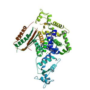
| Title | Crystal Structure of Heterocyst Differentiation Protein, HetR from Fischerella mv11 |
| Authors | Kim, Y., Joachimiak, G., Ye, Z., Binkowski, T.A., Zhang, R., Gornicki, P., Callahan, S.M., Hess, W.R., Haselkorn, R., Joachimiak, A. |
| Bioentity | None |
| R / Rfree | 0.20 / 0.27 |
| Unit cell edges [Å] | 123.38 x 123.38 x 109.61 |
| Unit cell angles [°] | 90.0, 90.0, 120.0 |
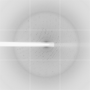
Dataset wih10su2-peak1.####.img details
| Number of frames | 136 (1 - 136) |
| Distance [mm] | 420.0 |
| Oscillation width [°] | 0.75 |
| Phi [°] | -120.0 |
| Wavelength [Å] | 0.97926 |
| Equipment | 19-ID at APS (Advanced Photon Source) |
Dataset wih10su2-peak2.####.img details
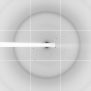
| Number of frames | 120 (1 - 120) |
| Distance [mm] | 420.0 |
| Oscillation width [°] | 0.75 |
| Phi [°] | -30.0 |
| Wavelength [Å] | 0.97926 |
| Equipment | 19-ID at APS (Advanced Photon Source) |