Diffraction project datasets SP19007A_3qec
Project details
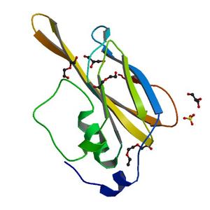
| Title |
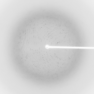
Crystal structure of a putative carbohydrate binding protein (PA1324) from Pseudomonas aeruginosa at 2.61 A resolution |
| Authors |
Joint Center for Structural Genomics (JCSG) |
| Bioentity |
None |
| R / Rfree |
0.21 / 0.23 |
| Unit cell edges [Å] |
132.79 x
132.79 x
132.79
|
| Unit cell angles [°] |
90.0,
90.0,
90.0
|
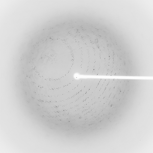
Dataset 163636_1_E1_####.mccd details
| Number of frames |
90 (361 - 450) |
| Distance [mm] |
370.0 |
| Oscillation width [°] |
0.50 |
| Phi [°] |
102.0 |
| Wavelength [Å] |
0.98012 |
| Experiment Date |
2010-11-23 |
| Equipment |
BL11-1
at SSRL (Stanford Synchrotron Radiation Laboratory)
|
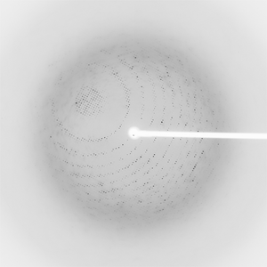
Dataset 163636_1_E2_####.mccd details
| Number of frames |
90 (361 - 450) |
| Distance [mm] |
370.0 |
| Oscillation width [°] |
0.50 |
| Phi [°] |
102.0 |
| Wavelength [Å] |
0.91837 |
| Experiment Date |
2010-11-23 |
| Equipment |
BL11-1
at SSRL (Stanford Synchrotron Radiation Laboratory)
|
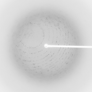
Dataset 163636_2_####.mccd details
| Number of frames |
90 (361 - 450) |
| Distance [mm] |
370.0 |
| Oscillation width [°] |
0.50 |
| Phi [°] |
102.0 |
| Wavelength [Å] |
0.97944 |
| Experiment Date |
2010-11-23 |
| Equipment |
BL11-1
at SSRL (Stanford Synchrotron Radiation Laboratory)
|
Dataset 163636_3_####.mccd details
| Number of frames |
90 (361 - 450) |
| Distance [mm] |
370.0 |
| Oscillation width [°] |
0.50 |
| Phi [°] |
147.0 |
| Wavelength [Å] |
0.97944 |
| Experiment Date |
2010-11-23 |
| Equipment |
BL11-1
at SSRL (Stanford Synchrotron Radiation Laboratory)
|