Diffraction project datasets SP17965A_3noh
Project details
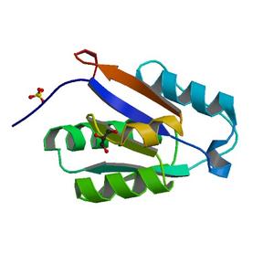
| Title |
Crystal structure of a putative peptide binding protein (RUMGNA_00914) from Ruminococcus gnavus ATCC 29149 at 1.60 A resolution |
| Authors |
Joint Center for Structural Genomics (JCSG) |
| Bioentity |
None |
| R / Rfree |
0.16 / 0.20 |
| Unit cell edges [Å] |
62.96 x
62.96 x
52.98
|
| Unit cell angles [°] |
90.0,
90.0,
120.0
|
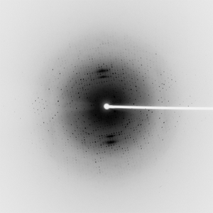
Dataset 156999_1_E1_####.mccd details
| Number of frames |
120 (1 - 120) |
| Distance [mm] |
200.0 |
| Oscillation width [°] |
1.00 |
| Phi [°] |
295.0 |
| Wavelength [Å] |
0.97917 |
| Experiment Date |
2010-06-10 |
| Equipment |
BL11-1
at SSRL (Stanford Synchrotron Radiation Laboratory)
|
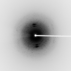
Dataset 156999_1_E2_####.mccd details
| Number of frames |
120 (1 - 120) |
| Distance [mm] |
200.0 |
| Oscillation width [°] |
1.00 |
| Phi [°] |
295.0 |
| Wavelength [Å] |
0.91837 |
| Experiment Date |
2010-06-10 |
| Equipment |
BL11-1
at SSRL (Stanford Synchrotron Radiation Laboratory)
|
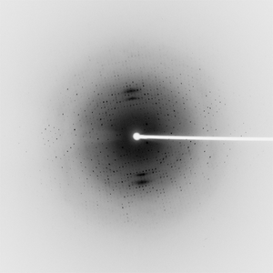
Dataset 156999_2_####.mccd details
| Number of frames |
120 (1 - 120) |
| Distance [mm] |
200.0 |
| Oscillation width [°] |
1.00 |
| Phi [°] |
295.0 |
| Wavelength [Å] |
0.97874 |
| Experiment Date |
2010-06-10 |
| Equipment |
BL11-1
at SSRL (Stanford Synchrotron Radiation Laboratory)
|