Diffraction project datasets SP17657A_3npd
- Method: MAD
- Resolution: 1.6 Å
- Space group: P 21 21 21
PDB website for 3NPD
PDB validation report for 3NPD
doi:10.18430/M33NPD
Project details
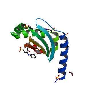
| Title | Crystal structure of a putative secreted protein (PA3611) from PSEUDOMONAS AERUGINOSA at 1.60 A resolution |
| Authors | Das, D., Chiu, H.J., Farr, C.L., Grant, J.C., Jaroszewski, L., Knuth, M.W., Miller, M.D., Tien, H.J., Elsliger, M.A., Deacon, A.M., Godzik, A., Lesley, S.A., Wilson, I.A. |
| Bioentity | None |
| R / Rfree | 0.16 / 0.21 |
| Unit cell edges [Å] | 34.99 x 51.32 x 56.90 |
| Unit cell angles [°] | 90.0, 90.0, 90.0 |
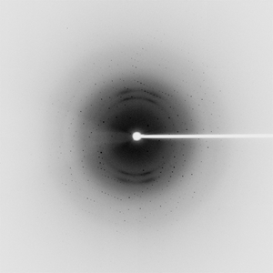
Dataset 153348_1_E1_####.mccd details
| Number of frames | 90 (1 - 90) |
| Distance [mm] | 200.0 |
| Oscillation width [°] | 1.00 |
| Phi [°] | 124.0 |
| Wavelength [Å] | 0.97941 |
| Experiment Date | 2010-05-12 |
| Equipment | BL9-2 at SSRL (Stanford Synchrotron Radiation Laboratory) |
Dataset 153348_1_E2_####.mccd details
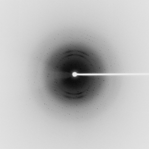
| Number of frames | 90 (1 - 90) |
| Distance [mm] | 200.0 |
| Oscillation width [°] | 1.00 |
| Phi [°] | 124.0 |
| Wavelength [Å] | 0.91837 |
| Experiment Date | 2010-05-12 |
| Equipment | BL9-2 at SSRL (Stanford Synchrotron Radiation Laboratory) |
Dataset 153348_2_####.mccd details
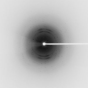
| Number of frames | 90 (1 - 90) |
| Distance [mm] | 200.0 |
| Oscillation width [°] | 1.00 |
| Phi [°] | 124.0 |
| Wavelength [Å] | 0.97925 |
| Experiment Date | 2010-05-12 |
| Equipment | BL9-2 at SSRL (Stanford Synchrotron Radiation Laboratory) |