Diffraction project datasets PS04222_3f7c
Project details
| Title |
Crystal structure of a duf416 family protein (maqu_0942) from marinobacter aquaeolei vt8 at 2.00 A resolution |
| Authors |
Joint Center for Structural Genomics (JCSG) |
| Bioentity |
None |
| R / Rfree |
0.17 / 0.20 |
| Unit cell edges [Å] |
77.00 x
77.00 x
91.26
|
| Unit cell angles [°] |
90.0,
90.0,
120.0
|
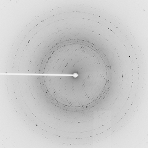
Dataset 97004_1_E1.#### details
| Number of frames |
90 (1 - 90) |
| Distance [mm] |
250.0 |
| Oscillation width [°] |
1.00 |
| Phi [°] |
-43.0 |
| Wavelength [Å] |
0.97967 |
| Experiment Date |
2008-10-12 |
| Equipment |
23-ID-B
at APS (Advanced Photon Source)
|
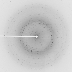
Dataset 97004_1_E2.#### details
| Number of frames |
90 (1 - 90) |
| Distance [mm] |
250.0 |
| Oscillation width [°] |
1.00 |
| Phi [°] |
-43.0 |
| Wavelength [Å] |
0.94645 |
| Experiment Date |
2008-10-12 |
| Equipment |
23-ID-B
at APS (Advanced Photon Source)
|
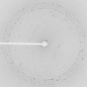
Dataset 97004_2.#### details
| Number of frames |
45 (1 - 45) |
| Distance [mm] |
500.0 |
| Oscillation width [°] |
2.00 |
| Phi [°] |
-43.0 |
| Wavelength [Å] |
0.94645 |
| Experiment Date |
2008-10-12 |
| Equipment |
23-ID-B
at APS (Advanced Photon Source)
|