Diffraction project datasets PK10409D_3go5
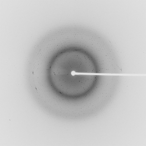
- Method: SAD
- Resolution: 1.4 Å
- Space group: P 32 2 1
PDB website for 3GO5
PDB validation report for 3GO5
doi:10.18430/M33GO5
Project details
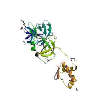
| Title | Crystal structure of a multidomain protein with nucleic acid binding domains (sp_0946) from streptococcus pneumoniae tigr4 at 1.40 A resolution |
| Authors | Matsumoto, Y., Xu, Q., Miyazaki, S., Kaito, C., Farr, C.L., Axelrod, H.L., Chiu, H.J., Klock, H.E., Knuth, M.W., Miller, M.D., Elsliger, M.A., Deacon, A.M., Godzik, A., Lesley, S.A., Sekimizu, K., Wilson, I.A. |
| Bioentity | None |
| R / Rfree | 0.15 / 0.18 |
| Unit cell edges [Å] | 62.56 x 62.56 x 160.20 |
| Unit cell angles [°] | 90.0, 90.0, 120.0 |
Dataset 102992_1_###.mccd details
| Number of frames | 23 (1 - 23) |
| Distance [mm] | 190.0 |
| Oscillation width [°] | 0.50 |
| Phi [°] | 348.0 |
| Wavelength [Å] | 0.97839 |
| Experiment Date | 2008-11-15 |
| Equipment | BL11-1 at SSRL (Stanford Synchrotron Radiation Laboratory) |
Dataset 102992_2_###.mccd details
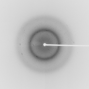
| Number of frames | 360 (1 - 360) |
| Distance [mm] | 190.0 |
| Oscillation width [°] | 0.50 |
| Phi [°] | 348.0 |
| Wavelength [Å] | 0.97839 |
| Experiment Date | 2008-11-15 |
| Equipment | BL11-1 at SSRL (Stanford Synchrotron Radiation Laboratory) |