Diffraction project datasets PC06304A_2o3l
Project details
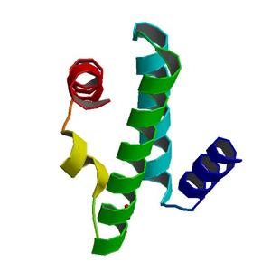
| Title |
Crystal structure of a duf1048 protein with a left-handed superhelix fold (bce_3448) from bacillus cereus atcc 10987 at 2.05 A resolution |
| Authors |
Joint Center for Structural Genomics (JCSG) |
| Bioentity |
None |
| R / Rfree |
0.18 / 0.23 |
| Unit cell edges [Å] |
61.20 x
61.20 x
101.70
|
| Unit cell angles [°] |
90.0,
90.0,
120.0
|
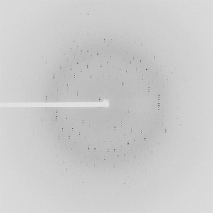
Dataset 34719_1_E1.#### details
| Number of frames |
90 (1 - 90) |
| Distance [mm] |
280.0 |
| Oscillation width [°] |
1.00 |
| Phi [°] |
114.0 |
| Wavelength [Å] |
0.97942 |
| Experiment Date |
2006-10-19 |
| Equipment |
23-ID-D
at APS (Advanced Photon Source)
|
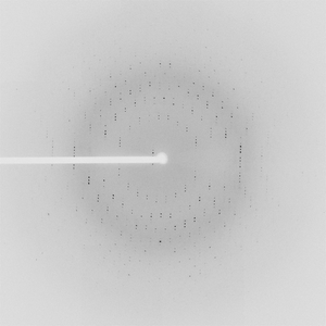
Dataset 34719_1_E2.#### details
| Number of frames |
90 (1 - 90) |
| Distance [mm] |
280.0 |
| Oscillation width [°] |
1.00 |
| Phi [°] |
114.0 |
| Wavelength [Å] |
0.94645 |
| Experiment Date |
2006-10-19 |
| Equipment |
23-ID-D
at APS (Advanced Photon Source)
|
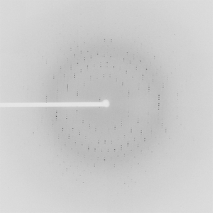
Dataset 34719_2.#### details
| Number of frames |
90 (1 - 90) |
| Distance [mm] |
280.0 |
| Oscillation width [°] |
1.00 |
| Phi [°] |
114.0 |
| Wavelength [Å] |
0.97921 |
| Experiment Date |
2006-10-19 |
| Equipment |
23-ID-D
at APS (Advanced Photon Source)
|