Diffraction project datasets PC05853B_3no5
Project details
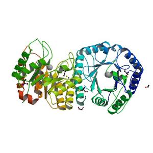
| Title |
Crystal structure of a Pfam DUF849 domain containing protein (Reut_A1631) from Ralstonia eutropha JMP134 at 1.90 A resolution |
| Authors |
Joint Center for Structural Genomics (JCSG) |
| Bioentity |
None |
| R / Rfree |
0.21 / 0.22 |
| Unit cell edges [Å] |
83.18 x
81.22 x
140.43
|
| Unit cell angles [°] |
90.0,
103.7,
90.0
|
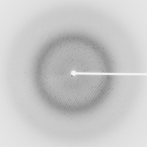
Dataset 42386_1_E1_###.mccd details
| Number of frames |
155 (1 - 155) |
| Distance [mm] |
275.0 |
| Oscillation width [°] |
1.00 |
| Phi [°] |
356.0 |
| Wavelength [Å] |
0.97921 |
| Experiment Date |
2007-02-11 |
| Equipment |
BL11-1
at SSRL (Stanford Synchrotron Radiation Laboratory)
|
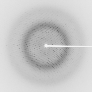
Dataset 42386_1_E2_###.mccd details
| Number of frames |
155 (1 - 155) |
| Distance [mm] |
275.0 |
| Oscillation width [°] |
1.00 |
| Phi [°] |
356.0 |
| Wavelength [Å] |
0.91162 |
| Experiment Date |
2007-02-11 |
| Equipment |
BL11-1
at SSRL (Stanford Synchrotron Radiation Laboratory)
|
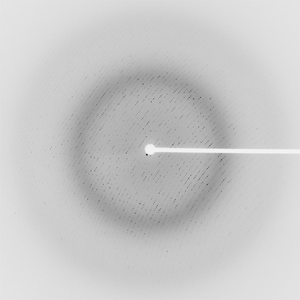
Dataset 42386_2_###.mccd details
| Number of frames |
155 (1 - 155) |
| Distance [mm] |
275.0 |
| Oscillation width [°] |
1.00 |
| Phi [°] |
356.0 |
| Wavelength [Å] |
0.97895 |
| Experiment Date |
2007-02-11 |
| Equipment |
BL11-1
at SSRL (Stanford Synchrotron Radiation Laboratory)
|