Diffraction project datasets MH7542A_3l12
Project details
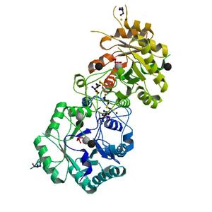
| Title |
Crystal structure of Putative glycerophosphoryl diester phosphodiesterase (YP_165505.1) from Silicibacter pomeroyi DSS-3 at 1.60 A resolution |
| Authors |
Joint Center for Structural Genomics (JCSG) |
| Bioentity |
None |
| R / Rfree |
0.16 / 0.19 |
| Unit cell edges [Å] |
50.41 x
136.41 x
50.55
|
| Unit cell angles [°] |
90.0,
118.0,
90.0
|
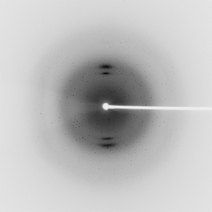
Dataset 125169_1_E1_###.mccd details
| Number of frames |
240 (1 - 240) |
| Distance [mm] |
220.0 |
| Oscillation width [°] |
0.50 |
| Phi [°] |
281.0 |
| Wavelength [Å] |
0.97917 |
| Experiment Date |
2009-06-12 |
| Equipment |
BL11-1
at SSRL (Stanford Synchrotron Radiation Laboratory)
|
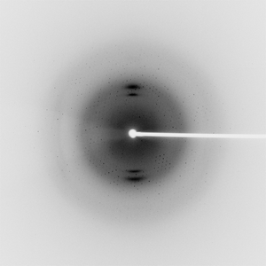
Dataset 125169_1_E2_###.mccd details
| Number of frames |
240 (1 - 240) |
| Distance [mm] |
220.0 |
| Oscillation width [°] |
0.50 |
| Phi [°] |
281.0 |
| Wavelength [Å] |
0.91837 |
| Experiment Date |
2009-06-12 |
| Equipment |
BL11-1
at SSRL (Stanford Synchrotron Radiation Laboratory)
|
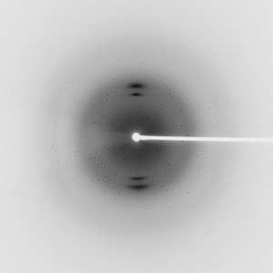
Dataset 125169_2_###.mccd details
| Number of frames |
240 (1 - 240) |
| Distance [mm] |
220.0 |
| Oscillation width [°] |
0.50 |
| Phi [°] |
281.0 |
| Wavelength [Å] |
0.97862 |
| Experiment Date |
2009-06-12 |
| Equipment |
BL11-1
at SSRL (Stanford Synchrotron Radiation Laboratory)
|