Diffraction project datasets MH4068H_3ks5
Project details
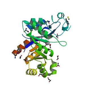
| Title |
Crystal structure of Putative glycerophosphoryl diester phosphodiesterase (17743486) from AGROBACTERIUM TUMEFACIENS str. C58 (Dupont) at 2.05 A resolution |
| Authors |
Joint Center for Structural Genomics (JCSG) |
| Bioentity |
None |
| R / Rfree |
0.21 / 0.24 |
| Unit cell edges [Å] |
84.15 x
84.15 x
214.88
|
| Unit cell angles [°] |
90.0,
90.0,
90.0
|
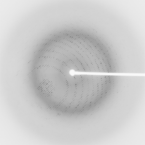
Dataset 118551_1_E1_###.mccd details
| Number of frames |
90 (1 - 90) |
| Distance [mm] |
300.0 |
| Oscillation width [°] |
1.00 |
| Phi [°] |
353.0 |
| Wavelength [Å] |
0.97855 |
| Experiment Date |
2009-04-15 |
| Equipment |
BL11-1
at SSRL (Stanford Synchrotron Radiation Laboratory)
|
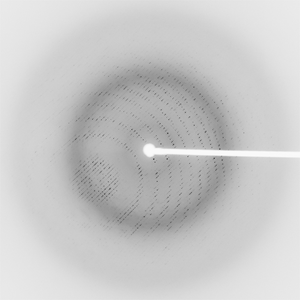
Dataset 118551_1_E2_###.mccd details
| Number of frames |
90 (1 - 90) |
| Distance [mm] |
300.0 |
| Oscillation width [°] |
1.00 |
| Phi [°] |
353.0 |
| Wavelength [Å] |
0.91837 |
| Experiment Date |
2009-04-15 |
| Equipment |
BL11-1
at SSRL (Stanford Synchrotron Radiation Laboratory)
|
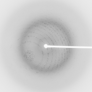
Dataset 118551_2_###.mccd details
| Number of frames |
90 (1 - 90) |
| Distance [mm] |
300.0 |
| Oscillation width [°] |
1.00 |
| Phi [°] |
353.0 |
| Wavelength [Å] |
0.97783 |
| Experiment Date |
2009-04-15 |
| Equipment |
BL11-1
at SSRL (Stanford Synchrotron Radiation Laboratory)
|