Diffraction project datasets IDP04384_3pzs
- Method: SAD
- Resolution: 1.89 Å
- Space group: P 21 21 21
PDB website for 3PZS
PDB validation report for 3PZS
doi:10.18430/m33pzs
Project details
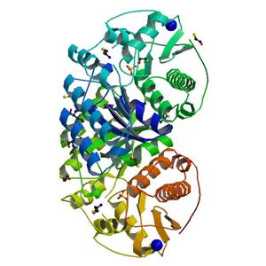
| Title | Crystal Structure of a pyridoxamine kinase from Yersinia pestis CO92 |
| Authors | Brunzelle, J.S., Wawrzak, Z., Kudritska, M., Onopriyenko, O., Savchenko, A., Anderson, W.F., Center for Structural Genomics of Infectious Diseases (CSGID) |
| R / Rfree | 0.17 / 0.20 |
| Unit cell edges [Å] | 67.91 x 88.10 x 102.92 |
| Unit cell angles [°] | 90.0, 90.0, 90.0 |
Dataset IDP04384-2.### details
| Number of frames | 320 (1 - 320) |
| Distance [mm] | 220.0 |
| Oscillation width [°] | 0.75 |
| Phi [°] | 145.0 |
| Wavelength [Å] | 0.97856 |
| Experiment Date | 2010-12-02 |
| Equipment | 21-ID-G at APS (Advanced Photon Source) |