Diffraction project datasets IDP04345_3o6v
- Method: Molecular Replacement
- Resolution: 1.695 Å
- Space group: H 3
PDB website for 3O6V
PDB validation report for 3O6V
doi:10.18430/m33o6v
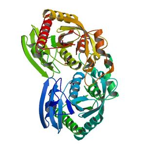
Project details
| Title | Crystal structure of Uridine Phosphorylase from Vibrio cholerae O1 biovar El Tor |
| Authors | Maltseva, N., Kim, Y., Hasseman, J., Anderson, W.F., Joachimiak, A., Center for Structural Genomics of Infectious Diseases (CSGID) |
| R / Rfree | 0.15 / 0.18 |
| Unit cell edges [Å] | 93.07 x 93.07 x 155.57 |
| Unit cell angles [°] | 90.0, 90.0, 120.0 |
Dataset IMPPEGsIIC9gly-peak.####.img details
| Number of frames | 58 (1 - 58) |
| Distance [mm] | 240.0 |
| Oscillation width [°] | 1.00 |
| Omega [°] | -100.0 |
| Kappa / Chi [°] | 0.0002 |
| Phi [°] | 89.9916 |
| Wavelength [Å] | 0.97921 |
| Experiment Date | 2010-04-23 |
| Equipment | 19-ID at APS (Advanced Photon Source) |
Dataset IMPPEGsIIC9gly-peak2.####.img details
| Number of frames | 120 (1 - 120) |
| Distance [mm] | 240.0 |
| Oscillation width [°] | 1.00 |
| Omega [°] | -60.0 |
| Kappa / Chi [°] | 0.0002 |
| Phi [°] | 89.9916 |
| Wavelength [Å] | 0.97921 |
| Experiment Date | 2010-04-23 |
| Equipment | 19-ID at APS (Advanced Photon Source) |