Diffraction project datasets IDP04069_3r3t
- Method: SAD
- Resolution: 2.302 Å
- Space group: P 31 2 1
PDB website for 3R3T
PDB validation report for 3R3T
doi:10.18430/m33r3t
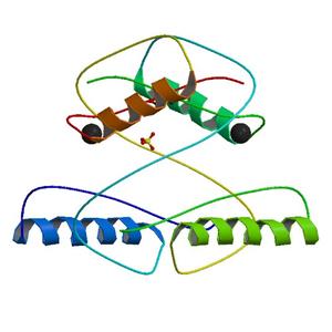
Project details
| Title | Crystal Structure of 30S Ribosomal Protein S from Bacillus anthracis |
| Authors | Kim, Y., Zhou, M., Kwon, K., Anderson, W.F., Joachimiak, A., Center for Structural Genomics of Infectious Diseases (CSGID) |
| R / Rfree | 0.21 / 0.23 |
| Unit cell edges [Å] | 82.68 x 82.68 x 59.17 |
| Unit cell angles [°] | 90.0, 90.0, 120.0 |
Dataset opt-i56c1-peak.####.img details
| Number of frames | 220 (1 - 220) |
| Distance [mm] | 330.0 |
| Oscillation width [°] | 1.00 |
| Omega [°] | -110.0 |
| Kappa / Chi [°] | 0.0001 |
| Phi [°] | -0.0037 |
| Wavelength [Å] | 0.97942 |
| Experiment Date | 2010-10-13 |
| Equipment | 19-ID at APS (Advanced Photon Source) |