Diffraction project datasets IDP02527_3l4e
- Method: SAD
- Resolution: 1.5 Å
- Space group: P 65 2 2
PDB website for 3L4E
PDB validation report for 3L4E
doi:10.18430/m33l4e
Project details
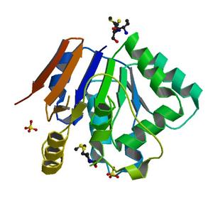
| Title | 1.5A Crystal Structure of a Putative Peptidase E Protein from Listeria monocytogenes EGD-e |
| Authors | Brunzelle, J.S., Onopriyenko, O., Savchenko, A., Anderson, W.F., Center for Structural Genomics of Infectious Diseases (CSGID) |
| R / Rfree | 0.17 / 0.22 |
| Unit cell edges [Å] | 67.82 x 67.82 x 155.17 |
| Unit cell angles [°] | 90.0, 90.0, 120.0 |
Dataset idp02527-1_A.### details
| Number of frames | 240 (1 - 240) |
| Distance [mm] | 180.0 |
| Oscillation width [°] | 0.75 |
| Phi [°] | 75.0 |
| Wavelength [Å] | 0.97857 |
| Experiment Date | 2009-11-12 |
| Equipment | 21-ID-D at APS (Advanced Photon Source) |
Dataset idp02527-1_B.### details
| Number of frames | 240 (1 - 240) |
| Distance [mm] | 180.0 |
| Oscillation width [°] | 0.75 |
| Phi [°] | 255.0 |
| Wavelength [Å] | 0.97857 |
| Experiment Date | 2009-11-12 |
| Equipment | 21-ID-D at APS (Advanced Photon Source) |