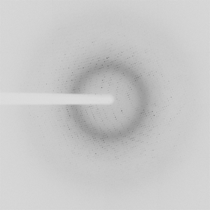
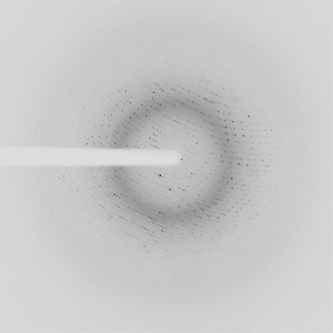
Diffraction project datasets IDP01937_5T1P

- Resolution: 2.0 Å
- Space group: P 1
Uniprot website for A0A0W8LFT3
PDB website for 5T1P
PDB validation report for 5T1P
doi:10.18430/m35t1p
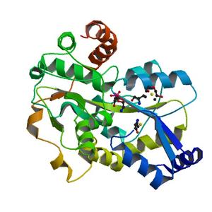
Project details
| Title | Crystal structure of the putative periplasmic solute-binding protein from Campylobacter jejuni |
| Authors | Filippova, E.V., Wawrzsak, Z., Sandoval, J., Skarina, T., Grimshaw, S., Savchenko, A., Anderson, W.F., Center for Structural Genomics of Infectious Diseases (CSGID) |
| R / Rfree | 0.18 / 0.22 |
| Unit cell edges [Å] | 82.19 x 89.28 x 100.39 |
| Unit cell angles [°] | 68.9, 82.5, 70.7 |
Dataset IDP01937-3.### details
| Number of frames | 777 (1 - 777) |
| Distance [mm] | 240.0 |
| Oscillation width [°] | 1.00 |
| Wavelength [Å] | 0.97856 |
| Experiment Date | 2016-04-14 |
| Equipment | 21-ID-G at APS (Advanced Photon Source) |
Dataset IDP01937-3b.### details
| Number of frames | 150 (1 - 150) |
| Distance [mm] | 240.0 |
| Oscillation width [°] | 1.00 |
| Wavelength [Å] | 0.97856 |
| Experiment Date | 2016-04-14 |
| Equipment | 21-ID-G at APS (Advanced Photon Source) |
Dataset IDP01937-3c.### details
| Number of frames | 165 (152 - 316) |
| Distance [mm] | 240.0 |
| Oscillation width [°] | 1.00 |
| Phi [°] | 151.0 |
| Wavelength [Å] | 0.97856 |
| Experiment Date | 2016-04-14 |
| Equipment | 21-ID-G at APS (Advanced Photon Source) |