Diffraction project datasets IDP01112_3g48
- Method: Molecular Replacement
- Resolution: 1.5 Å
- Space group: P 21 21 21
PDB website for 3G48
PDB validation report for 3G48
doi:10.18430/m33g48
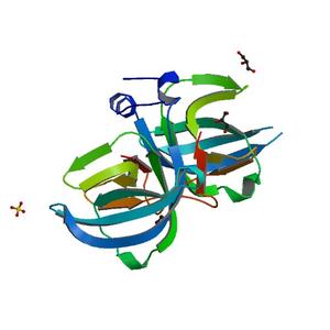
Project details
| Title | Crystal structure of chaperone CsaA form Bacillus anthracis str. Ames |
| Authors | Nocek, B., Zhou, M., Stam, J., Anderson, W., Joachimiak, A., Center for Structural Genomics of Infectious Diseases (CSGID) |
| R / Rfree | 0.17 / 0.20 |
| Unit cell edges [Å] | 39.36 x 67.56 x 74.64 |
| Unit cell angles [°] | 90.0, 90.0, 90.0 |
Dataset xh7.####.img details
| Number of frames | 311 (1 - 311) |
| Distance [mm] | 201.0 |
| Oscillation width [°] | 0.50 |
| Omega [°] | -88.0 |
| Kappa / Chi [°] | 0.0003 |
| Phi [°] | -0.007 |
| Wavelength [Å] | 0.97926 |
| Experiment Date | 2008-12-06 |
| Equipment | 19-ID at APS (Advanced Photon Source) |