Diffraction project datasets IDP00692_3h5q
- Method: SAD
- Resolution: 1.94 Å
- Space group: P 1 2 1
PDB website for 3H5Q
PDB validation report for 3H5Q
doi:10.18430/M33H5Q
Project details
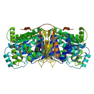
| Title | Crystal structure of a putative pyrimidine-nucleoside phosphorylase from Staphylococcus aureus |
| Authors | Shumilin, I.A., Zimmerman, M., Cymborowski, M., Skarina, T., Onopriyenko, O., Anderson, W.F., Savchenko, A., Minor, W., Center for Structural Genomics of Infectious Diseases (CSGID) |
| R / Rfree | 0.17 / 0.20 |
| Unit cell edges [Å] | 51.59 x 40.89 x 105.58 |
| Unit cell angles [°] | 90.0, 97.2, 90.0 |
Dataset IDP00692_ct1-1_####.img details
| Number of frames | 180 (1 - 180) |
| Distance [mm] | 290.0 |
| Oscillation width [°] | 1.00 |
| Omega [°] | -115.0 |
| Kappa / Chi [°] | 0.0003 |
| Phi [°] | -0.007 |
| Wavelength [Å] | 0.97857 |
| Experiment Date | 2008-11-21 |
| Equipment | 19-ID at APS (Advanced Photon Source) |