Diffraction project datasets IDP00456_3gsd
- Method: Molecular Replacement
- Resolution: 2.05 Å
- Space group: C 2 2 21
PDB website for 3GSD
PDB validation report for 3GSD
doi:10.18430/m33gsd
Project details
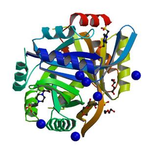
| Title | 2.05 Angstrom structure of a divalent-cation tolerance protein (CutA) from Yersinia pestis |
| Authors | Minasov, G., Wawrzak, Z., Skarina, T., Onopriyenko, O., Peterson, S.N., Savchenko, A., Anderson, W.F., Center for Structural Genomics of Infectious Diseases (CSGID) |
| R / Rfree | 0.16 / 0.20 |
| Unit cell edges [Å] | 140.78 x 157.91 x 157.13 |
| Unit cell angles [°] | 90.0, 90.0, 90.0 |
Dataset ypo346-3.### details
| Number of frames | 300 (1 - 300) |
| Distance [mm] | 220.0 |
| Oscillation width [°] | 0.50 |
| Phi [°] | 85.0 |
| Wavelength [Å] | 0.97872 |
| Experiment Date | 2009-02-27 |
| Equipment | 21-ID-F at APS (Advanced Photon Source) |